 Movie
Movie Controller


+ Open data

- Basic information
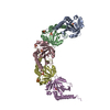
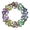
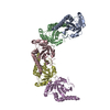
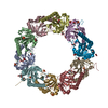
| Entry | Database: PDB / ID: 1e2y | ||||||
|---|---|---|---|---|---|---|---|
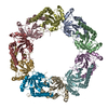
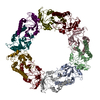
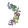
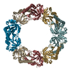
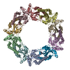
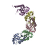
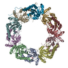
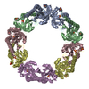
| Title | Tryparedoxin peroxidase from Crithidia fasciculata | ||||||
 Components Components | TRYPAREDOXIN PEROXIDASE | ||||||
 Keywords Keywords | OXIDOREDUCTASE / 2-CYS PEROXIREDOXIN | ||||||
| Function / homology |  Function and homology information Function and homology informationthioredoxin-dependent peroxiredoxin / thioredoxin peroxidase activity / cellular response to stress / cell redox homeostasis / hydrogen peroxide catabolic process / response to oxidative stress / cytosol Similarity search - Function | ||||||
| Biological species |  CRITHIDIA FASCICULATA (eukaryote) CRITHIDIA FASCICULATA (eukaryote) | ||||||
| Method |  X-RAY DIFFRACTION / SYNCHROTRON / MAD / Resolution: 3.2 Å X-RAY DIFFRACTION / SYNCHROTRON / MAD / Resolution: 3.2 Å | ||||||
 Authors Authors | Alphey, M.S. / Bond, C.S. / Hunter, W.N. | ||||||
 Citation Citation | Journal: J.Mol.Biol. / Year: 2000 Title: The Structure of Reduced Tryparedoxin Peroxidase Reveals a Decamer and Insight Into Reactivity of 2Cys-Peroxiredoxins Authors: Alphey, M.S. / Bond, C.S. / Tetaud, E. / Fairlamb, A.H. / Hunter, W.N. | ||||||
| History |
|
- Structure visualization
| Structure viewer | Molecule: MolmilJmol/JSmol |
|---|
- Downloads & links
-Download
| PDBx/mmCIF format | 1e2y.cif.gz | 320.5 KB | Display | PDBx/mmCIF format |
|---|---|---|---|---|
| PDB format | pdb1e2y.ent.gz | 265.6 KB | Display | PDB format |
| PDBx/mmJSON format | 1e2y.json.gz | Tree view | PDBx/mmJSON format | |
| Others |  Other downloads Other downloads |
-Validation report
| Arichive directory | https://data.pdbj.org/pub/pdb/validation_reports/e2/1e2yftp://data.pdbj.org/pub/pdb/validation_reports/e2/1e2y | HTTPS FTP |
|---|
-Related structure data
| Similar structure data |
|---|


-Links
 PDBj
PDBj- Assembly
| Deposited unit | 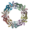
| ||||||||||||||||||||
|---|---|---|---|---|---|---|---|---|---|---|---|---|---|---|---|---|---|---|---|---|---|
| 1 |
| ||||||||||||||||||||
| Unit cell |
| ||||||||||||||||||||
| Noncrystallographic symmetry (NCS) | NCS oper:
|
-Components
| #1: Protein | Mass: 21313.393 Da / Num. of mol.: 10 Source method: isolated from a genetically manipulated source Source: (gene. exp.) CRITHIDIA FASCICULATA (eukaryote)Description: PLASMID TRANSFORMED INTO E. COLI BL21(DE3) STRAIN FOR RECOMBINANT PROTEIN EXPRESSION Cellular location: CYTOPLASM / Gene: CF-TRYP / Production host:  #2: Chemical | ChemComp-CL /   Mass: 35.453 Da / Num. of mol.: 10 / Source method: obtained synthetically / Formula: Cl Mass: 35.453 Da / Num. of mol.: 10 / Source method: obtained synthetically / Formula: Cl#3: Water | ChemComp-HOH / |  Mass: 18.015 Da / Num. of mol.: 22 / Source method: isolated from a natural source / Formula: H2O Mass: 18.015 Da / Num. of mol.: 22 / Source method: isolated from a natural source / Formula: H2OHas protein modification | Y | |
|---|
-Experimental details
-Experiment
| Experiment | Method: X-RAY DIFFRACTION / Number of used crystals: 1 |
|---|
- Sample preparation
| Crystal | Density Matthews: 3.5 Å3/Da / Density % sol: 65 % | ||||||||||||||||||||||||||||||||||||||||
|---|---|---|---|---|---|---|---|---|---|---|---|---|---|---|---|---|---|---|---|---|---|---|---|---|---|---|---|---|---|---|---|---|---|---|---|---|---|---|---|---|---|
| Crystal grow | Method: vapor diffusion, hanging drop / pH: 7.5 Details: PROTEIN CRYSTALLIZED FROM 12.5% PEG 1000, 100MM TRIS-HCL PH HANGING DROP CONSISITED | ||||||||||||||||||||||||||||||||||||||||
| Crystal grow | *PLUS Method: vapor diffusion, hanging dropDetails: drop consists of 2.5 micro litter of protein mixed with 2 micro litter of reservoir solution and 0.5 micro litter EDTA | ||||||||||||||||||||||||||||||||||||||||
| Components of the solutions | *PLUS
|
-Data collection
| Diffraction | Mean temperature: 100 K | ||||||||||||
|---|---|---|---|---|---|---|---|---|---|---|---|---|---|
| Diffraction source | Source: SYNCHROTRON / Site: ESRF  / Beamline: BM14 / Wavelength: 0.8800,0.9790,0.9792 / Beamline: BM14 / Wavelength: 0.8800,0.9790,0.9792 | ||||||||||||
| Detector | Type: MARRESEARCH / Detector: CCD / Date: Jun 15, 1999 / Details: MIRROR | ||||||||||||
| Radiation | Monochromator: SI(111) / Protocol: MAD / Monochromatic (M) / Laue (L): M / Scattering type: x-ray | ||||||||||||
| Radiation wavelength |
| ||||||||||||
| Reflection | Resolution: 3.2→21 Å / Num. obs: 52094 / % possible obs: 99.8 % / Redundancy: 4 % / Biso Wilson estimate: 56.4 Å2 / Rsym value: 0.091 / Net I/σ(I): 14.8 | ||||||||||||
| Reflection shell | Resolution: 3.2→21 Å / Redundancy: 4 % / Mean I/σ(I) obs: 2.6 / Rsym value: 0.48 / % possible all: 96.7 | ||||||||||||
| Reflection | *PLUS Num. measured all: 376643 / Rmerge(I) obs: 0.091 | ||||||||||||
| Reflection shell | *PLUS % possible obs: 96.7 % / Rmerge(I) obs: 0.48 |
- Processing
| Software |
| ||||||||||||||||||||||||||||||||||||||||||||||||||||||||||||
|---|---|---|---|---|---|---|---|---|---|---|---|---|---|---|---|---|---|---|---|---|---|---|---|---|---|---|---|---|---|---|---|---|---|---|---|---|---|---|---|---|---|---|---|---|---|---|---|---|---|---|---|---|---|---|---|---|---|---|---|---|---|
| Refinement | Method to determine structure: MAD / Resolution: 3.2→21 Å / Rfactor Rfree error: 0.006 / Data cutoff high absF: 10000 / Cross valid method: THROUGHOUT / σ(F): 0
| ||||||||||||||||||||||||||||||||||||||||||||||||||||||||||||
| Solvent computation | Solvent model: DENSITY MODIFICATION / Bsol: 12.362 Å2 / ksol: 0.205 e/Å3 | ||||||||||||||||||||||||||||||||||||||||||||||||||||||||||||
| Displacement parameters | Biso mean: 71 Å2
| ||||||||||||||||||||||||||||||||||||||||||||||||||||||||||||
| Refine analyze |
| ||||||||||||||||||||||||||||||||||||||||||||||||||||||||||||
| Refinement step | Cycle: LAST / Resolution: 3.2→21 Å
| ||||||||||||||||||||||||||||||||||||||||||||||||||||||||||||
| Refine LS restraints |
| ||||||||||||||||||||||||||||||||||||||||||||||||||||||||||||
| Refine LS restraints NCS | NCS model details: RESTRAINTS | ||||||||||||||||||||||||||||||||||||||||||||||||||||||||||||
| LS refinement shell | Resolution: 3.2→3.31 Å / Rfactor Rfree error: 0.036 / Total num. of bins used: 10
| ||||||||||||||||||||||||||||||||||||||||||||||||||||||||||||
| Software | *PLUS Name: CNS / Version: 1 / Classification: refinement | ||||||||||||||||||||||||||||||||||||||||||||||||||||||||||||
| Refine LS restraints | *PLUS
|
