 Movie
Movie Controller


+ Open data

- Basic information
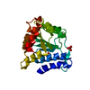
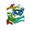
| Entry | Database: PDB / ID: 2ewf | ||||||
|---|---|---|---|---|---|---|---|
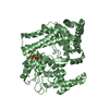
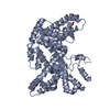
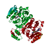
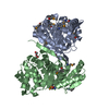
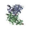
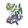
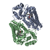
| Title | Crystal structure of the site-specific DNA nickase N.BspD6I | ||||||
 Components Components | Nicking endonuclease N.BspD6I | ||||||
 Keywords Keywords | HYDROLASE / helix-turn-helix / beta-alpha-barrel | ||||||
| Function / homology |  Function and homology information Function and homology information | ||||||
| Biological species |  | ||||||
| Method |  X-RAY DIFFRACTION / SYNCHROTRON / MAD / Resolution: 1.84 Å X-RAY DIFFRACTION / SYNCHROTRON / MAD / Resolution: 1.84 Å | ||||||
 Authors Authors | Kachalova, G.S. / Bartunik, H.D. / Artyukh, R.I. / Rogulin, E.A. / Perevyazova, T.A. / Zheleznaya, L.A. / Matvienko, N.I. | ||||||
 Citation Citation | Journal: J.Mol.Biol. / Year: 2008 Title: Structural analysis of the heterodimeric type IIS restriction endonuclease R.BspD6I acting as a complex between a monomeric site-specific nickase and a catalytic subunit. Authors: Kachalova, G.S. / Rogulin, E.A. / Yunusova, A.K. / Artyukh, R.I. / Perevyazova, T.A. / Matvienko, N.I. / Zheleznaya, L.A. / Bartunik, H.D. #1: Journal: Acta Crystallogr.,Sect.F / Year: 2005Title: Crystallization and preliminary crystallographic analysis of the site-specific DNA nickase Nb.BspD6I Authors: Kachalova, G.S. / Rogulin, E.A. / Artyukh, R.I. / Perevyazova, T.A. / Zheleznaya, L.A. / Matvienko, N.I. / Bartunik, H.D. #2: Journal: Biochemistry Mosc. / Year: 2003Title: Cloning and sequencing of the gene of site-specific nickase N.BspD6I Authors: Perevyazova, T.A. / Rogulin, E.A. / Zheleznaya, L.A. / Matvienko, N.I. | ||||||
| History |
|
- Structure visualization
| Structure viewer | Molecule: MolmilJmol/JSmol |
|---|
- Downloads & links
-Download
| PDBx/mmCIF format | 2ewf.cif.gz | 149.7 KB | Display | PDBx/mmCIF format |
|---|---|---|---|---|
| PDB format | pdb2ewf.ent.gz | 115 KB | Display | PDB format |
| PDBx/mmJSON format | 2ewf.json.gz | Tree view | PDBx/mmJSON format | |
| Others |  Other downloads Other downloads |
-Validation report
| Arichive directory | https://data.pdbj.org/pub/pdb/validation_reports/ew/2ewfftp://data.pdbj.org/pub/pdb/validation_reports/ew/2ewf | HTTPS FTP |
|---|
-Related structure data
-Links
 PDBj
PDBj

- Assembly
| Deposited unit | 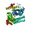
| ||||||||
|---|---|---|---|---|---|---|---|---|---|
| 1 |
| ||||||||
| Unit cell |
|
-Components
| #1: Protein | Mass: 71702.078 Da / Num. of mol.: 1 Source method: isolated from a genetically manipulated source Source: (gene. exp.) | ||
|---|---|---|---|
| #2: Chemical | ChemComp-BR /   Mass: 79.904 Da / Num. of mol.: 13 / Source method: obtained synthetically / Formula: Br Mass: 79.904 Da / Num. of mol.: 13 / Source method: obtained synthetically / Formula: Br#3: Water | ChemComp-HOH / |  Mass: 18.015 Da / Num. of mol.: 627 / Source method: isolated from a natural source / Formula: H2O Mass: 18.015 Da / Num. of mol.: 627 / Source method: isolated from a natural source / Formula: H2O |
-Experimental details
-Experiment
| Experiment | Method: X-RAY DIFFRACTION / Number of used crystals: 1 |
|---|
- Sample preparation
| Crystal | Density Matthews: 2.72 Å3/Da / Density % sol: 54.82 % |
|---|---|
| Crystal grow | Temperature: 298 K / Method: vapor diffusion, sitting drop / pH: 7.2 Details: 0.04 M KH2PO4, 16% (w/v) PEG 8000, 20% (v/v) glycerol, pH 7.2, VAPOR DIFFUSION, SITTING DROP, temperature 298.0K |
-Data collection
| Diffraction | Mean temperature: 100 K | ||||||||||||
|---|---|---|---|---|---|---|---|---|---|---|---|---|---|
| Diffraction source | Source: SYNCHROTRON / Site: MPG/DESY, HAMBURG  / Beamline: BW6 / Wavelength: 0.9198, 0.9200, 1.05 / Beamline: BW6 / Wavelength: 0.9198, 0.9200, 1.05 | ||||||||||||
| Detector | Type: MARRESEARCH / Detector: CCD / Date: Jul 25, 2005 | ||||||||||||
| Radiation | Monochromator: graphite / Protocol: MAD / Monochromatic (M) / Laue (L): M / Scattering type: x-ray | ||||||||||||
| Radiation wavelength |
| ||||||||||||
| Reflection | Resolution: 1.84→20 Å / Num. obs: 67523 / % possible obs: 99.9 % / Observed criterion σ(F): 2 / Observed criterion σ(I): 2 / Rmerge(I) obs: 0.08 / Net I/σ(I): 12.6 | ||||||||||||
| Reflection shell | Resolution: 1.84→1.87 Å / % possible all: 100 |
- Processing
| Software |
| |||||||||||||||||||||||||||||||||
|---|---|---|---|---|---|---|---|---|---|---|---|---|---|---|---|---|---|---|---|---|---|---|---|---|---|---|---|---|---|---|---|---|---|---|
| Refinement | Method to determine structure: MAD / Resolution: 1.84→10 Å / Num. parameters: 22116 / Num. restraintsaints: 20045 / Cross valid method: FREE R / σ(F): 0 / Stereochemistry target values: ENGH AND HUBER / Details: ANISOTROPIC REFINEMENT
| |||||||||||||||||||||||||||||||||
| Refine analyze | Num. disordered residues: 7 / Occupancy sum hydrogen: 0 / Occupancy sum non hydrogen: 5358.79
| |||||||||||||||||||||||||||||||||
| Refinement step | Cycle: LAST / Resolution: 1.84→10 Å
| |||||||||||||||||||||||||||||||||
| Refine LS restraints |
| |||||||||||||||||||||||||||||||||
| LS refinement shell | Resolution: 1.84→1.87 Å / Redundancy reflection obs: 3131
|
