 Movie
Movie Controller

[English] 日本語
 Yorodumi
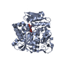
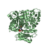
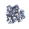
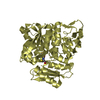
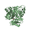
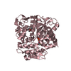
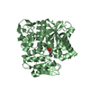
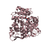
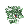
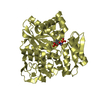
- PDB-5b3a: Crystal Structure of O-Phoshoserine Sulfhydrylase from Aeropyrum ... -
+ Open data

- Basic information
| Entry | Database: PDB / ID: 5b3a | ||||||
|---|---|---|---|---|---|---|---|
| Title | Crystal Structure of O-Phoshoserine Sulfhydrylase from Aeropyrum pernix in Complexed with the alpha-Aminoacrylate Intermediate | ||||||
 Components Components | Protein CysO | ||||||
 Keywords Keywords | TRANSFERASE / Cysteine Biosynthesis / sulfhydrylase / intermediate / External Schiff base of PLP with alpha-amino acrylate | ||||||
| Function / homology |  Function and homology information Function and homology informationO-phosphoserine sulfhydrylase / O-phosphoserine sulfhydrylase activity / cystathionine beta-synthase / cystathionine beta-synthase activity / cysteine synthase / cysteine synthase activity / : Similarity search - Function | ||||||
| Biological species |   Aeropyrum pernix K1 (archaea) Aeropyrum pernix K1 (archaea) | ||||||
| Method |  X-RAY DIFFRACTION / SYNCHROTRON / FOURIER SYNTHESIS / Resolution: 2.14 Å X-RAY DIFFRACTION / SYNCHROTRON / FOURIER SYNTHESIS / Resolution: 2.14 Å | ||||||
 Authors Authors | Nakamura, T. / Takeda, E. / Kawai, Y. / Kataoka, M. / Ishikawa, K. | ||||||
 Citation Citation | Journal: Extremophiles / Year: 2016 Title: Role of F225 in O-phosphoserine sulfhydrylase from Aeropyrum pernix K1 Authors: Takeda, E. / Kunimoto, K. / Kawai, Y. / Kataoka, M. / Ishikawa, K. / Nakamura, T. #1: Journal: J.Mol.Biol. / Year: 2012Title: Structural analysis of the substrate recognition mechanism in O-phosphoserine sulfhydrylase from the hyperthermophilic archaeon Aeropyrum pernix K1 Authors: Nakamura, T. / Kawai, Y. / Kunimoto, K. / Iwasaki, Y. / Nishii, K. / Kataoka, M. / Ishikawa, K. #2: Journal: J.Mol.Biol. / Year: 2005Title: Three-dimensional structure of a new enzyme, O-phosphoserine sulfhydrylase, involved in l-cysteine biosynthesis by a hyperthermophilic archaeon, Aeropyrum pernix K1, at 2.0A resolution Authors: Oda, Y. / Mino, K. / Ishikawa, K. / Ataka, M. | ||||||
| History |
|
- Structure visualization
| Structure viewer | Molecule: MolmilJmol/JSmol |
|---|
- Downloads & links
-Download
| PDBx/mmCIF format | 5b3a.cif.gz | 167.2 KB | Display | PDBx/mmCIF format |
|---|---|---|---|---|
| PDB format | pdb5b3a.ent.gz | 131.6 KB | Display | PDB format |
| PDBx/mmJSON format | 5b3a.json.gz | Tree view | PDBx/mmJSON format | |
| Others |  Other downloads Other downloads |
-Validation report
| Arichive directory | https://data.pdbj.org/pub/pdb/validation_reports/b3/5b3aftp://data.pdbj.org/pub/pdb/validation_reports/b3/5b3a | HTTPS FTP |
|---|
-Related structure data
| Related structure data | 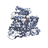 5b36C 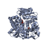 3vsaS C: citing same article ( S: Starting model for refinement |
|---|---|
| Similar structure data |


-Links
 PDBj
PDBj
- Assembly
| Deposited unit | 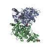
| ||||||||
|---|---|---|---|---|---|---|---|---|---|
| 1 | 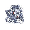
| ||||||||
| 2 | 
| ||||||||
| Unit cell |
|
-Components
| #1: Protein | Mass: 42025.855 Da / Num. of mol.: 2 Source method: isolated from a genetically manipulated source Source: (gene. exp.) Aeropyrum pernix K1 (archaea) / Strain: K1 / Gene: cysO, APE_1586 / Plasmid: pET3d / Production host:  References: UniProt: Q9YBL2, cystathionine beta-synthase, cysteine synthase, O-phosphoserine sulfhydrylase #2: Chemical |   Mass: 118.174 Da / Num. of mol.: 2 / Source method: obtained synthetically / Formula: C6H14O2 / Comment: precipitant*YM Mass: 118.174 Da / Num. of mol.: 2 / Source method: obtained synthetically / Formula: C6H14O2 / Comment: precipitant*YM#3: Chemical | 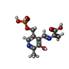  Mass: 316.204 Da / Num. of mol.: 2 / Source method: obtained synthetically / Formula: C11H13N2O7P Mass: 316.204 Da / Num. of mol.: 2 / Source method: obtained synthetically / Formula: C11H13N2O7P#4: Water | ChemComp-HOH / |  Mass: 18.015 Da / Num. of mol.: 348 / Source method: isolated from a natural source / Formula: H2O Mass: 18.015 Da / Num. of mol.: 348 / Source method: isolated from a natural source / Formula: H2O |
|---|
-Experimental details
-Experiment
| Experiment | Method: X-RAY DIFFRACTION / Number of used crystals: 1 |
|---|
- Sample preparation
| Crystal | Density Matthews: 2.26 Å3/Da / Density % sol: 45.49 % |
|---|---|
| Crystal grow | Temperature: 296 K / Method: vapor diffusion, hanging drop / pH: 7.5 Details: 0.1 M HEPES sodium pH 7.5, 27% 2-propanol, 12% PEG4000, 12 mM TCEP-HCl, the crystal was soaked with the reservoir solution containing 5% MPD as a cryoprotectant, 20 mM O-phospho-L-serine (OPS) and 12 mM TCEP-HCl |
-Data collection
| Diffraction | Mean temperature: 80 K |
|---|---|
| Diffraction source | Source: SYNCHROTRON / Site: SPring-8  / Beamline: BL44XU / Wavelength: 0.9 Å / Beamline: BL44XU / Wavelength: 0.9 Å |
| Detector | Type: Bruker DIP-6040 / Detector: CCD / Date: Nov 24, 2009 |
| Radiation | Monochromator: Rotated-inclined double-crystal monochromator , Si (111) Protocol: SINGLE WAVELENGTH / Monochromatic (M) / Laue (L): M / Scattering type: x-ray |
| Radiation wavelength | Wavelength: 0.9 Å / Relative weight: 1 |
| Reflection | Resolution: 2.14→32.92 Å / Num. obs: 40639 / % possible obs: 97.99 % / Redundancy: 10 % / Net I/σ(I): 59.8 |
| Reflection shell | Resolution: 2.14→2.18 Å |
- Processing
| Software |
| ||||||||||||||||||||||||||||||||||||||||||||||||||||||||||||||||||||||||||||||||||||||||||||||||||||||||||||||||||||||||||||||||||||||||||||||||||||||||||||||||||||||||||||||||||||||
|---|---|---|---|---|---|---|---|---|---|---|---|---|---|---|---|---|---|---|---|---|---|---|---|---|---|---|---|---|---|---|---|---|---|---|---|---|---|---|---|---|---|---|---|---|---|---|---|---|---|---|---|---|---|---|---|---|---|---|---|---|---|---|---|---|---|---|---|---|---|---|---|---|---|---|---|---|---|---|---|---|---|---|---|---|---|---|---|---|---|---|---|---|---|---|---|---|---|---|---|---|---|---|---|---|---|---|---|---|---|---|---|---|---|---|---|---|---|---|---|---|---|---|---|---|---|---|---|---|---|---|---|---|---|---|---|---|---|---|---|---|---|---|---|---|---|---|---|---|---|---|---|---|---|---|---|---|---|---|---|---|---|---|---|---|---|---|---|---|---|---|---|---|---|---|---|---|---|---|---|---|---|---|---|
| Refinement | Method to determine structure: FOURIER SYNTHESIS Starting model: CysO free form (PDB ID, 3VSA) Resolution: 2.14→32.92 Å / Cor.coef. Fo:Fc: 0.962 / Cor.coef. Fo:Fc free: 0.93 / SU B: 3.821 / SU ML: 0.101 / Cross valid method: THROUGHOUT / ESU R: 0.2 / ESU R Free: 0.176 / Details: HYDROGENS HAVE BEEN ADDED IN THE RIDING POSITIONS
| ||||||||||||||||||||||||||||||||||||||||||||||||||||||||||||||||||||||||||||||||||||||||||||||||||||||||||||||||||||||||||||||||||||||||||||||||||||||||||||||||||||||||||||||||||||||
| Solvent computation | Ion probe radii: 0.8 Å / Shrinkage radii: 0.8 Å / VDW probe radii: 1.2 Å | ||||||||||||||||||||||||||||||||||||||||||||||||||||||||||||||||||||||||||||||||||||||||||||||||||||||||||||||||||||||||||||||||||||||||||||||||||||||||||||||||||||||||||||||||||||||
| Displacement parameters | Biso mean: 19.593 Å2
| ||||||||||||||||||||||||||||||||||||||||||||||||||||||||||||||||||||||||||||||||||||||||||||||||||||||||||||||||||||||||||||||||||||||||||||||||||||||||||||||||||||||||||||||||||||||
| Refinement step | Cycle: 1 / Resolution: 2.14→32.92 Å
| ||||||||||||||||||||||||||||||||||||||||||||||||||||||||||||||||||||||||||||||||||||||||||||||||||||||||||||||||||||||||||||||||||||||||||||||||||||||||||||||||||||||||||||||||||||||
| Refine LS restraints |
|