 Movie
Movie Controller

[English] 日本語
 Yorodumi
- PDB-6l0q: Crystal Structure of the O-Phosphoserine Sulfhydrylase from Aerop... -
+ Open data

- Basic information
| Entry | Database: PDB / ID: 6l0q | ||||||
|---|---|---|---|---|---|---|---|
| Title | Crystal Structure of the O-Phosphoserine Sulfhydrylase from Aeropyrum pernix Complexed with O-Phosphoserine | ||||||
 Components Components | Protein CysO | ||||||
 Keywords Keywords | TRANSFERASE / CYSTEINE BIOSYNTHESIS / SULFHYDRYLASE | ||||||
| Function / homology |  Function and homology information Function and homology informationO-phosphoserine sulfhydrylase / O-phosphoserine sulfhydrylase activity / cystathionine beta-synthase / cystathionine beta-synthase activity / cysteine synthase / cysteine synthase activity / : Similarity search - Function | ||||||
| Biological species |   Aeropyrum pernix K1 (archaea) Aeropyrum pernix K1 (archaea) | ||||||
| Method |  X-RAY DIFFRACTION / SYNCHROTRON / MOLECULAR REPLACEMENT / molecular replacement / Resolution: 1.58 Å X-RAY DIFFRACTION / SYNCHROTRON / MOLECULAR REPLACEMENT / molecular replacement / Resolution: 1.58 Å | ||||||
 Authors Authors | Nakabayashi, M. / Takeda, E. / Ishikawa, K. / Nakamura, T. | ||||||
 Citation Citation | Journal: J.Biosci.Bioeng. / Year: 2021 Title: Identification of amino acid residues important for recognition of O-phospho-l-serine substrates by cysteine synthase. Authors: Takeda, E. / Matsui, E. / Kiryu, T. / Nakagawa, T. / Nakabayashi, M. / Ishikawa, K. / Nakamura, T. | ||||||
| History |
|
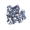
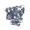
- Structure visualization
| Structure viewer | Molecule: MolmilJmol/JSmol |
|---|
- Downloads & links
-Download
| PDBx/mmCIF format | 6l0q.cif.gz | 303.2 KB | Display | PDBx/mmCIF format |
|---|---|---|---|---|
| PDB format | pdb6l0q.ent.gz | 245.6 KB | Display | PDB format |
| PDBx/mmJSON format | 6l0q.json.gz | Tree view | PDBx/mmJSON format | |
| Others |  Other downloads Other downloads |
-Validation report
| Arichive directory | https://data.pdbj.org/pub/pdb/validation_reports/l0/6l0qftp://data.pdbj.org/pub/pdb/validation_reports/l0/6l0q | HTTPS FTP |
|---|
-Related structure data
| Related structure data | 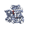 6l0pC 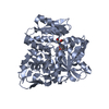 6l0rC 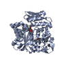 6l0sC 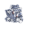 5b3aS S: Starting model for refinement C: citing same article ( |
|---|---|
| Similar structure data |


-Links
 PDBj
PDBj
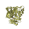
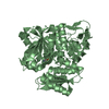
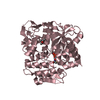
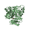
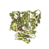
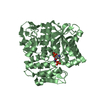
- Assembly
| Deposited unit | 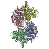
| ||||||||
|---|---|---|---|---|---|---|---|---|---|
| 1 | 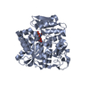
| ||||||||
| 2 | 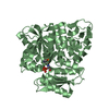
| ||||||||
| 3 | 
| ||||||||
| 4 | 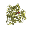
| ||||||||
| Unit cell |
|
-Components
| #1: Protein | Mass: 41983.754 Da / Num. of mol.: 4 / Mutation: K127A, F225Y Source method: isolated from a genetically manipulated source Source: (gene. exp.) Aeropyrum pernix K1 (archaea) / Strain: K1 / Gene: cysO, APE_1586 / Plasmid: pET3D / Production host:  References: UniProt: Q9YBL2, cystathionine beta-synthase, cysteine synthase, O-phosphoserine sulfhydrylase #2: Chemical | ChemComp-E1U / (   Mass: 414.199 Da / Num. of mol.: 4 / Source method: obtained synthetically / Formula: C11H16N2O11P2 / Feature type: SUBJECT OF INVESTIGATION Mass: 414.199 Da / Num. of mol.: 4 / Source method: obtained synthetically / Formula: C11H16N2O11P2 / Feature type: SUBJECT OF INVESTIGATION#3: Chemical |   Mass: 118.174 Da / Num. of mol.: 2 / Source method: obtained synthetically / Formula: C6H14O2 / Comment: precipitant*YM Mass: 118.174 Da / Num. of mol.: 2 / Source method: obtained synthetically / Formula: C6H14O2 / Comment: precipitant*YM#4: Water | ChemComp-HOH / |  Mass: 18.015 Da / Num. of mol.: 305 / Source method: isolated from a natural source / Formula: H2O Mass: 18.015 Da / Num. of mol.: 305 / Source method: isolated from a natural source / Formula: H2OHas ligand of interest | Y | |
|---|
-Experimental details
-Experiment
| Experiment | Method: X-RAY DIFFRACTION / Number of used crystals: 1 |
|---|
- Sample preparation
| Crystal | Density Matthews: 2.32 Å3/Da / Density % sol: 46.92 % |
|---|---|
| Crystal grow | Temperature: 296 K / Method: vapor diffusion, hanging drop Details: 0.1M sodium N-2-hydroxyethylpiperazine-N'-2-ethanesulfonate buffer, pH 8.2 (7.9), 29%(v/v) 2-propanol, 13% (11%) (v/v) polyethylene glycol 4,000, and 11mM TCEP-HCl |
-Data collection
| Diffraction | Mean temperature: 100 K / Serial crystal experiment: N |
|---|---|
| Diffraction source | Source: SYNCHROTRON / Site: SPring-8  / Beamline: BL44XU / Wavelength: 0.9 Å / Beamline: BL44XU / Wavelength: 0.9 Å |
| Detector | Type: Bruker DIP-6040 / Detector: CCD / Date: Oct 29, 2013 |
| Radiation | Protocol: SINGLE WAVELENGTH / Monochromatic (M) / Laue (L): M / Scattering type: x-ray |
| Radiation wavelength | Wavelength: 0.9 Å / Relative weight: 1 |
| Reflection | Resolution: 1.58→75.2 Å / Num. obs: 208050 / % possible obs: 100 % / Redundancy: 7.5 % / Rmerge(I) obs: 0.133 / Net I/σ(I): 7.5 |
| Reflection shell | Resolution: 1.58→1.66 Å / Rmerge(I) obs: 1.038 / Mean I/σ(I) obs: 1.7 / Num. unique obs: 30464 |
-Phasing
| Phasing | Method: molecular replacement |
|---|
- Processing
| Software |
| ||||||||||||||||||||||||||||||||||||||||||||||||||||||||||||
|---|---|---|---|---|---|---|---|---|---|---|---|---|---|---|---|---|---|---|---|---|---|---|---|---|---|---|---|---|---|---|---|---|---|---|---|---|---|---|---|---|---|---|---|---|---|---|---|---|---|---|---|---|---|---|---|---|---|---|---|---|---|
| Refinement | Method to determine structure: MOLECULAR REPLACEMENT Starting model: 5B3A Resolution: 1.58→75.2 Å / Cor.coef. Fo:Fc: 0.968 / Cor.coef. Fo:Fc free: 0.965 / WRfactor Rfree: 0.1859 / WRfactor Rwork: 0.1801 / FOM work R set: 0.9229 / SU B: 0.822 / SU ML: 0.031 / SU R Cruickshank DPI: 0.0764 / SU Rfree: 0.0695 / Cross valid method: THROUGHOUT / σ(F): 0 / ESU R: 0.076 / ESU R Free: 0.069 / Stereochemistry target values: MAXIMUM LIKELIHOOD Details: HYDROGENS HAVE BEEN ADDED IN THE RIDING POSITIONS U VALUES : REFINED INDIVIDUALLY
| ||||||||||||||||||||||||||||||||||||||||||||||||||||||||||||
| Solvent computation | Ion probe radii: 0.8 Å / Shrinkage radii: 0.8 Å / VDW probe radii: 1.2 Å / Solvent model: MASK | ||||||||||||||||||||||||||||||||||||||||||||||||||||||||||||
| Displacement parameters | Biso max: 68.91 Å2 / Biso mean: 24.685 Å2 / Biso min: 9.34 Å2
| ||||||||||||||||||||||||||||||||||||||||||||||||||||||||||||
| Refinement step | Cycle: final / Resolution: 1.58→75.2 Å
| ||||||||||||||||||||||||||||||||||||||||||||||||||||||||||||
| Refine LS restraints |
| ||||||||||||||||||||||||||||||||||||||||||||||||||||||||||||
| LS refinement shell | Resolution: 1.58→1.62 Å / Rfactor Rfree error: 0
|
