- PDB-5l95: Crystal structure of human UBA5 in complex with UFM1 and AMP -
+
Open data
ID or keywords:
Loading...
-
Basic information
Entry
Database: PDB / ID: 5l95
Title
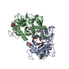
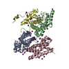
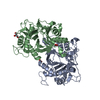
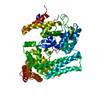
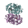
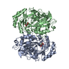
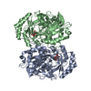
Crystal structure of human UBA5 in complex with UFM1 and AMP
Components
Ubiquitin-fold modifier 1
Ubiquitin-like modifier-activating enzyme 5
Keywords
CELL CYCLE / Ubiquitin like protein E1 / UBL
Function / homology
Function and homology information
UFM1 activating enzyme activity / protein K69-linked ufmylation / protein ufmylation / megakaryocyte differentiation / regulation of type II interferon production / regulation of intracellular estrogen receptor signaling pathway / reticulophagy / negative regulation of protein import into nucleus / neuromuscular process / response to endoplasmic reticulum stress ...UFM1 activating enzyme activity / protein K69-linked ufmylation / protein ufmylation / megakaryocyte differentiation / regulation of type II interferon production / regulation of intracellular estrogen receptor signaling pathway / reticulophagy / negative regulation of protein import into nucleus / neuromuscular process / response to endoplasmic reticulum stress / erythrocyte differentiation / brain development / Antigen processing: Ubiquitination & Proteasome degradation / endoplasmic reticulum membrane / negative regulation of apoptotic process / endoplasmic reticulum / Golgi apparatus / protein homodimerization activity / zinc ion binding / ATP binding / nucleus / cytoplasm / cytosol Similarity search - Function
Ubiquitin-fold modifier 1 / Ubiquitin fold modifier 1 protein / D-isomer specific 2-hydroxyacid dehydrogenase, NAD-binding domain conserved site 1 / ThiF/MoeB/HesA family / THIF-type NAD/FAD binding fold / ThiF family / Ubiquitin-activating enzyme / Phosphatidylinositol 3-kinase Catalytic Subunit; Chain A, domain 1 / Ubiquitin-like (UB roll) / NAD(P)-binding Rossmann-like Domain ...Ubiquitin-fold modifier 1 / Ubiquitin fold modifier 1 protein / D-isomer specific 2-hydroxyacid dehydrogenase, NAD-binding domain conserved site 1 / ThiF/MoeB/HesA family / THIF-type NAD/FAD binding fold / ThiF family / Ubiquitin-activating enzyme / Phosphatidylinositol 3-kinase Catalytic Subunit; Chain A, domain 1 / Ubiquitin-like (UB roll) / NAD(P)-binding Rossmann-like Domain / Ubiquitin-like domain superfamily / Roll / Rossmann fold / 3-Layer(aba) Sandwich / Alpha Beta Similarity search - Domain/homology
In the structure databanks used in Yorodumi, some data are registered as the other names, "COVID-19 virus" and "2019-nCoV". Here are the details of the virus and the list of structure data.
Jan 31, 2019. EMDB accession codes are about to change! (news from PDBe EMDB page)
EMDB accession codes are about to change! (news from PDBe EMDB page)
The allocation of 4 digits for EMDB accession codes will soon come to an end. Whilst these codes will remain in use, new EMDB accession codes will include an additional digit and will expand incrementally as the available range of codes is exhausted. The current 4-digit format prefixed with “EMD-” (i.e. EMD-XXXX) will advance to a 5-digit format (i.e. EMD-XXXXX), and so on. It is currently estimated that the 4-digit codes will be depleted around Spring 2019, at which point the 5-digit format will come into force.
The EM Navigator/Yorodumi systems omit the EMD- prefix.
Related info.:Q: What is EMD? / ID/Accession-code notation in Yorodumi/EM Navigator
Yorodumi is a browser for structure data from EMDB, PDB, SASBDB, etc.
This page is also the successor to EM Navigator detail page, and also detail information page/front-end page for Omokage search.
The word "yorodu" (or yorozu) is an old Japanese word meaning "ten thousand". "mi" (miru) is to see.
Related info.:EMDB / PDB / SASBDB / Comparison of 3 databanks / Yorodumi Search / Aug 31, 2016. New EM Navigator & Yorodumi / Yorodumi Papers / Jmol/JSmol / Function and homology information / Changes in new EM Navigator and Yorodumi
 Movie
Movie Controller


 Open data
 Basic information
 Components
 Keywords
 Function and homology information
 Homo sapiens (human)
 X-RAY DIFFRACTION /
  Authors
 Israel, 1items
  Citation
 Structure visualization
 Downloads & links
 Other downloads
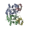


 PDBj
PDBj


 Assembly
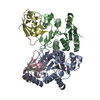


 Mass: 347.221 Da / Num. of mol.: 1 / Source method: obtained synthetically / Formula: C10H14N5O7P / Comment: AMP*YM
 Mass: 65.409 Da / Num. of mol.: 2 / Source method: obtained synthetically / Formula: Zn
 Mass: 18.015 Da / Num. of mol.: 193 / Source method: isolated from a natural source / Formula: H2O
 Sample preparation
 / Beamline: I03 / Wavelength: 0.97949 Å
 Processing