[English] 日本語
Yorodumi
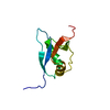
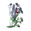
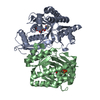
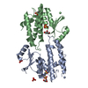
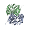
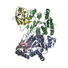
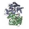
- PDB-5iaa: Crystal structure of human UBA5 in complex with UFM1 -

+
Open data


ID or keywords:

Loading...

-
Basic information

Entry
Database: PDB / ID: 5iaa
TitleCrystal structure of human UBA5 in complex with UFM1
Components
  • Ubiquitin-fold modifier 1
  • Ubiquitin-like modifier-activating enzyme 5
KeywordsCELL CYCLE / Ubiquitin like protein and E1
Function / homology
Function and homology information


UFM1 activating enzyme activity / protein ufmylation / protein K69-linked ufmylation / megakaryocyte differentiation / regulation of intracellular estrogen receptor signaling pathway / reticulophagy / neuromuscular process / negative regulation of protein import into nucleus / response to endoplasmic reticulum stress / erythrocyte differentiation ...UFM1 activating enzyme activity / protein ufmylation / protein K69-linked ufmylation / megakaryocyte differentiation / regulation of intracellular estrogen receptor signaling pathway / reticulophagy / neuromuscular process / negative regulation of protein import into nucleus / response to endoplasmic reticulum stress / erythrocyte differentiation / brain development / Antigen processing: Ubiquitination & Proteasome degradation / intracellular membrane-bounded organelle / endoplasmic reticulum membrane / negative regulation of apoptotic process / Golgi apparatus / endoplasmic reticulum / protein homodimerization activity / zinc ion binding / ATP binding / nucleus / cytosol / cytoplasm
Similarity search - Function
Ubiquitin-fold modifier 1 / Ubiquitin fold modifier 1 protein / D-isomer specific 2-hydroxyacid dehydrogenase, NAD-binding domain conserved site 1 / ThiF/MoeB/HesA family / Ubiquitin-activating enzyme / THIF-type NAD/FAD binding fold / ThiF family / Phosphatidylinositol 3-kinase Catalytic Subunit; Chain A, domain 1 / Ubiquitin-like (UB roll) / NAD(P)-binding Rossmann-like Domain ...Ubiquitin-fold modifier 1 / Ubiquitin fold modifier 1 protein / D-isomer specific 2-hydroxyacid dehydrogenase, NAD-binding domain conserved site 1 / ThiF/MoeB/HesA family / Ubiquitin-activating enzyme / THIF-type NAD/FAD binding fold / ThiF family / Phosphatidylinositol 3-kinase Catalytic Subunit; Chain A, domain 1 / Ubiquitin-like (UB roll) / NAD(P)-binding Rossmann-like Domain / Ubiquitin-like domain superfamily / Roll / Rossmann fold / 3-Layer(aba) Sandwich / Alpha Beta
Similarity search - Domain/homology
Ubiquitin-fold modifier 1 / Ubiquitin-like modifier-activating enzyme 5
Similarity search - Component
Biological speciesHomo sapiens (human)
MethodX-RAY DIFFRACTION / SYNCHROTRON / MOLECULAR REPLACEMENT / Resolution: 1.85 Å
AuthorsOweis, W. / Padala, P. / Wiener, R.
Funding support Israel, 1items
OrganizationGrant numberCountry
BSF Israel
CitationJournal: Cell Rep / Year: 2016
Title: Trans-Binding Mechanism of Ubiquitin-like Protein Activation Revealed by a UBA5-UFM1 Complex.
Authors: Oweis, W. / Padala, P. / Hassouna, F. / Cohen-Kfir, E. / Gibbs, D.R. / Todd, E.A. / Berndsen, C.E. / Wiener, R.
History
DepositionFeb 21, 2016Deposition site: PDBE / Processing site: PDBE
Revision 1.0Sep 28, 2016Provider: repository / Type: Initial release
Revision 1.1Oct 5, 2016Group: Database references
Revision 1.2Jan 10, 2024Group: Data collection / Database references / Refinement description
Category: chem_comp_atom / chem_comp_bond ...chem_comp_atom / chem_comp_bond / database_2 / pdbx_initial_refinement_model / struct_ncs_dom_lim
Item: _database_2.pdbx_DOI / _database_2.pdbx_database_accession ..._database_2.pdbx_DOI / _database_2.pdbx_database_accession / _struct_ncs_dom_lim.beg_auth_comp_id / _struct_ncs_dom_lim.beg_label_asym_id / _struct_ncs_dom_lim.beg_label_comp_id / _struct_ncs_dom_lim.beg_label_seq_id / _struct_ncs_dom_lim.end_auth_comp_id / _struct_ncs_dom_lim.end_label_asym_id / _struct_ncs_dom_lim.end_label_comp_id / _struct_ncs_dom_lim.end_label_seq_id

-
Structure visualization

Structure viewerMolecule:
MolmilJmol/JSmol

Downloads & links

-
Assembly

Deposited unit
A: Ubiquitin-like modifier-activating enzyme 5
B: Ubiquitin-like modifier-activating enzyme 5
C: Ubiquitin-fold modifier 1
D: Ubiquitin-fold modifier 1
hetero molecules


Theoretical massNumber of molelcules
Total (without water)82,4056
Polymers82,2744
Non-polymers1312
Water2,630146
1


  • Idetical with deposited unit
  • defined by software
TypeNameSymmetry operationNumber
identity operation1_555x,y,z1
Buried area10450 Å2
ΔGint-61 kcal/mol
Surface area27240 Å2
MethodPISA
Unit cell
Length a, b, c (Å)138.081, 138.081, 99.178
Angle α, β, γ (deg.)90.00, 90.00, 120.00
Int Tables number154
Space group name H-MP3221
Noncrystallographic symmetry (NCS)NCS domain:
IDEns-IDDetails
11A
21B
12C
22D

NCS domain segments:

Component-ID: _ / Refine code: _

Dom-IDEns-IDBeg auth comp-IDBeg label comp-IDEnd auth comp-IDEnd label comp-IDAuth asym-IDLabel asym-IDAuth seq-IDLabel seq-ID
11TYRTYRVALVALAA68 - 34612 - 290
21TYRTYRVALVALBB68 - 34612 - 290
12VALVALGLYGLYCC4 - 834 - 83
22VALVALGLYGLYDD4 - 834 - 83

NCS ensembles :
ID
1
2

-
Components

#1: Protein Ubiquitin-like modifier-activating enzyme 5 / Ubiquitin-activating enzyme 5 / ThiFP1 / UFM1-activating enzyme / Ubiquitin-activating enzyme E1 ...Ubiquitin-activating enzyme 5 / ThiFP1 / UFM1-activating enzyme / Ubiquitin-activating enzyme E1 domain-containing protein 1


Mass: 32198.885 Da / Num. of mol.: 2 / Fragment: UNP residues 57-346
Source method: isolated from a genetically manipulated source
Source: (gene. exp.) Homo sapiens (human) / Gene: UBA5, UBE1DC1 / Production host: Escherichia coli (E. coli) / References: UniProt: Q9GZZ9
#2: Protein Ubiquitin-fold modifier 1


Mass: 8938.331 Da / Num. of mol.: 2
Source method: isolated from a genetically manipulated source
Source: (gene. exp.) Homo sapiens (human) / Gene: UFM1, C13orf20, BM-002 / Production host: Escherichia coli (E. coli) / References: UniProt: P61960
#3: Chemical ChemComp-ZN / ZINC ION


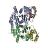
Mass: 65.409 Da / Num. of mol.: 2 / Source method: obtained synthetically / Formula: Zn
#4: Water ChemComp-HOH / water


Mass: 18.015 Da / Num. of mol.: 146 / Source method: isolated from a natural source / Formula: H2O

-
Experimental details

-
Experiment

ExperimentMethod: X-RAY DIFFRACTION / Number of used crystals: 1

-
Sample preparation

CrystalDensity Matthews: 3.32 Å3/Da / Density % sol: 62.92 %
Crystal growTemperature: 293.15 K / Method: vapor diffusion, hanging drop / Details: 8% Tascimate, pH 7.0 and 16% PEG3350

-
Data collection

DiffractionMean temperature: 100 K
Diffraction sourceSource: SYNCHROTRON / Site: BESSY / Beamline: 14.3 / Wavelength: 0.896 Å
DetectorType: DECTRIS PILATUS 6M / Detector: PIXEL / Date: Aug 25, 2015
RadiationProtocol: SINGLE WAVELENGTH / Monochromatic (M) / Laue (L): M / Scattering type: x-ray
Radiation wavelengthWavelength: 0.896 Å / Relative weight: 1
Reflection twin
Crystal-IDIDOperatorDomain-IDFraction
11H, K, L10.871
11-h,-k,l20.129
ReflectionResolution: 1.85→76.3 Å / Num. obs: 93040 / % possible obs: 100 % / Redundancy: 18.5 % / Rmerge(I) obs: 0.174 / Net I/σ(I): 11.6
Reflection shellHighest resolution: 1.85 Å

-
Processing

Software
NameVersionClassification
REFMAC5.8.0135refinement
iMOSFLMdata reduction
SCALAdata scaling
PHASERphasing
RefinementMethod to determine structure: MOLECULAR REPLACEMENT
Starting model: 1WXS and 3H8V
Resolution: 1.85→69.04 Å / Cor.coef. Fo:Fc: 0.962 / Cor.coef. Fo:Fc free: 0.952 / SU B: 3.987 / SU ML: 0.06 / Cross valid method: THROUGHOUT / ESU R: 0.021 / ESU R Free: 0.02 / Stereochemistry target values: MAXIMUM LIKELIHOOD / Details: HYDROGENS HAVE BEEN ADDED IN THE RIDING POSITIONS
RfactorNum. reflection% reflectionSelection details
Rfree0.20605 4641 5 %RANDOM
Rwork0.18741 ---
obs0.18834 88360 99.99 %-
Solvent computationIon probe radii: 0.7 Å / Shrinkage radii: 0.7 Å / VDW probe radii: 1 Å / Solvent model: MASK
Displacement parametersBiso mean: 42.487 Å2
Baniso -1Baniso -2Baniso -3
1-29.33 Å20 Å20 Å2
2--29.33 Å20 Å2
3----58.66 Å2
Refinement stepCycle: LAST / Resolution: 1.85→69.04 Å
ProteinNucleic acidLigandSolventTotal
Num. atoms5070 0 2 146 5218
Refine LS restraints
Refine-IDTypeDev idealDev ideal targetNumber
X-RAY DIFFRACTIONr_bond_refined_d0.0150.0195180
X-RAY DIFFRACTIONr_bond_other_d0.0070.024986
X-RAY DIFFRACTIONr_angle_refined_deg1.7661.9637023
X-RAY DIFFRACTIONr_angle_other_deg1.3933.00111462
X-RAY DIFFRACTIONr_dihedral_angle_1_deg6.4895650
X-RAY DIFFRACTIONr_dihedral_angle_2_deg35.96725.088226
X-RAY DIFFRACTIONr_dihedral_angle_3_deg13.92515865
X-RAY DIFFRACTIONr_dihedral_angle_4_deg15.4281522
X-RAY DIFFRACTIONr_chiral_restr0.1020.2812
X-RAY DIFFRACTIONr_gen_planes_refined0.010.0215866
X-RAY DIFFRACTIONr_gen_planes_other0.0050.021142
X-RAY DIFFRACTIONr_nbd_refined
X-RAY DIFFRACTIONr_nbd_other
X-RAY DIFFRACTIONr_nbtor_refined
X-RAY DIFFRACTIONr_nbtor_other
X-RAY DIFFRACTIONr_xyhbond_nbd_refined
X-RAY DIFFRACTIONr_xyhbond_nbd_other
X-RAY DIFFRACTIONr_metal_ion_refined
X-RAY DIFFRACTIONr_metal_ion_other
X-RAY DIFFRACTIONr_symmetry_vdw_refined
X-RAY DIFFRACTIONr_symmetry_vdw_other
X-RAY DIFFRACTIONr_symmetry_hbond_refined
X-RAY DIFFRACTIONr_symmetry_hbond_other
X-RAY DIFFRACTIONr_symmetry_metal_ion_refined
X-RAY DIFFRACTIONr_symmetry_metal_ion_other
X-RAY DIFFRACTIONr_mcbond_it1.5991.1962630
X-RAY DIFFRACTIONr_mcbond_other1.5991.1962629
X-RAY DIFFRACTIONr_mcangle_it2.4071.7723270
X-RAY DIFFRACTIONr_mcangle_other2.4071.7723271
X-RAY DIFFRACTIONr_scbond_it2.5131.4292549
X-RAY DIFFRACTIONr_scbond_other2.5131.4292549
X-RAY DIFFRACTIONr_scangle_it
X-RAY DIFFRACTIONr_scangle_other3.3662.0663753
X-RAY DIFFRACTIONr_long_range_B_refined6.73710.2735699
X-RAY DIFFRACTIONr_long_range_B_other6.73310.0725663
X-RAY DIFFRACTIONr_rigid_bond_restr
X-RAY DIFFRACTIONr_sphericity_free
X-RAY DIFFRACTIONr_sphericity_bonded
Refine LS restraints NCS

Refine-ID: X-RAY DIFFRACTION / Type: interatomic distance / Weight position: 0.05

Ens-IDDom-IDAuth asym-IDNumberRms dev position (Å)
11A274180.1
12B274180.1
21C90460.09
22D90460.09
LS refinement shellResolution: 1.85→1.898 Å / Total num. of bins used: 20
RfactorNum. reflection% reflection
Rfree0.373 323 -
Rwork0.353 6486 -
obs--99.99 %
Refinement TLS params.

Method: refined / Refine-ID: X-RAY DIFFRACTION

IDL112)L122)L132)L222)L232)L332)S11 (Å °)S12 (Å °)S13 (Å °)S21 (Å °)S22 (Å °)S23 (Å °)S31 (Å °)S32 (Å °)S33 (Å °)T112)T122)T132)T222)T232)T332)Origin x (Å)Origin y (Å)Origin z (Å)
10.5728-0.04190.01791.53-0.13472.25260.0141-0.0372-0.01940.0448-0.02680.0751-0.0422-0.09810.01270.13120.0650.00460.04030.00410.4924-75.71525.324-0.496
20.9326-0.0937-0.2111.7191-0.22032.01090.0267-0.0367-0.0493-0.0303-0.06820.11630.1631-0.04820.04150.22130.0624-0.01720.02950.00260.4936-74.773-3.931-6.923
31.4196-0.81190.04021.2492-1.62973.51350.3370.17950.0429-0.3132-0.11830.15120.0535-0.1439-0.21870.43280.2069-0.01940.1674-0.03840.5834-86.09329.354-24.202
41.7218-0.2388-1.44591.91710.64562.79-0.0268-0.0296-0.0147-0.07930.1938-0.2137-0.00810.432-0.1670.17250.0669-0.01990.1534-0.00120.5301-53.2960.586-20.582
Refinement TLS group
IDRefine-IDRefine TLS-IDAuth asym-IDAuth seq-ID
1X-RAY DIFFRACTION1A68 - 346
2X-RAY DIFFRACTION2B68 - 346
3X-RAY DIFFRACTION3C4 - 83
4X-RAY DIFFRACTION4D4 - 83

+
About Yorodumi

-
News

-
Feb 9, 2022. New format data for meta-information of EMDB entries

New format data for meta-information of EMDB entries

  • Version 3 of the EMDB header file is now the official format.
  • The previous official version 1.9 will be removed from the archive.

Related info.:EMDB header

External links:wwPDB to switch to version 3 of the EMDB data model

-
Aug 12, 2020. Covid-19 info

Covid-19 info

URL: https://pdbjlc1.pdbj.org/emnavi/covid19.php

New page: Covid-19 featured information page in EM Navigator.

Related info.:Covid-19 info / Mar 5, 2020. Novel coronavirus structure data

+
Mar 5, 2020. Novel coronavirus structure data

Novel coronavirus structure data

Related info.:Yorodumi Speices / Aug 12, 2020. Covid-19 info

External links:COVID-19 featured content - PDBj / Molecule of the Month (242):Coronavirus Proteases

+
Jan 31, 2019. EMDB accession codes are about to change! (news from PDBe EMDB page)

EMDB accession codes are about to change! (news from PDBe EMDB page)

  • The allocation of 4 digits for EMDB accession codes will soon come to an end. Whilst these codes will remain in use, new EMDB accession codes will include an additional digit and will expand incrementally as the available range of codes is exhausted. The current 4-digit format prefixed with “EMD-” (i.e. EMD-XXXX) will advance to a 5-digit format (i.e. EMD-XXXXX), and so on. It is currently estimated that the 4-digit codes will be depleted around Spring 2019, at which point the 5-digit format will come into force.
  • The EM Navigator/Yorodumi systems omit the EMD- prefix.

Related info.:Q: What is EMD? / ID/Accession-code notation in Yorodumi/EM Navigator

External links:EMDB Accession Codes are Changing Soon! / Contact to PDBj

+
Jul 12, 2017. Major update of PDB

Major update of PDB

  • wwPDB released updated PDB data conforming to the new PDBx/mmCIF dictionary.
  • This is a major update changing the version number from 4 to 5, and with Remediation, in which all the entries are updated.
  • In this update, many items about electron microscopy experimental information are reorganized (e.g. em_software).
  • Now, EM Navigator and Yorodumi are based on the updated data.

External links:wwPDB Remediation / Enriched Model Files Conforming to OneDep Data Standards Now Available in the PDB FTP Archive

-
Yorodumi

Thousand views of thousand structures

  • Yorodumi is a browser for structure data from EMDB, PDB, SASBDB, etc.
  • This page is also the successor to EM Navigator detail page, and also detail information page/front-end page for Omokage search.
  • The word "yorodu" (or yorozu) is an old Japanese word meaning "ten thousand". "mi" (miru) is to see.

Related info.:EMDB / PDB / SASBDB / Comparison of 3 databanks / Yorodumi Search / Aug 31, 2016. New EM Navigator & Yorodumi / Yorodumi Papers / Jmol/JSmol / Function and homology information / Changes in new EM Navigator and Yorodumi

Read more