Component-ID: 1 / Ens-ID: 1 / Beg auth comp-ID: ASN / Beg label comp-ID: ASN / End auth comp-ID: SER / End label comp-ID: SER / Refine code: 6 / Auth seq-ID: 3 - 358 / Label seq-ID: 4 - 359
Dom-ID
Auth asym-ID
Label asym-ID
1
A
A
2
B
B
Details
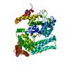
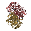
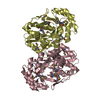
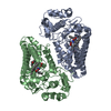
ANALYTICAL SIZE EXCLUSION CHROMATOGRAPHY WITH STATIC LIGHT SCATTERING SUPPORTS THE ASSIGNMENT OF A DIMER AS A SIGNIFICANT OLIGOMERIZATION STATE IN SOLUTION
-
Components
#1: Protein
AlcoholdehydrogenaseIV / Maleylacetate reductase
Mass: 38769.691 Da / Num. of mol.: 2 Source method: isolated from a genetically manipulated source Source: (gene. exp.) Corynebacterium glutamicum (bacteria) / Gene: cg3386, Cgl3057, NP_602249.1, tcbF / Plasmid: SpeedET / Production host: Escherichia coli (E. coli) / Strain (production host): HK100 / References: UniProt: Q8NL91, maleylacetate reductase
Mass: 18.015 Da / Num. of mol.: 397 / Source method: isolated from a natural source / Formula: H2O
Has protein modification
Y
Sequence details
THIS CONSTRUCT (RESIDUES 1-363) WAS EXPRESSED WITH A PURIFICATION TAG MGSDKIHHHHHHENLYFQG. THE TAG ...THIS CONSTRUCT (RESIDUES 1-363) WAS EXPRESSED WITH A PURIFICATION TAG MGSDKIHHHHHHENLYFQG. THE TAG WAS REMOVED WITH TEV PROTEASE LEAVING ONLY A GLYCINE (0) FOLLOWED BY THE TARGET SEQUENCE.
-
Experimental details
-
Experiment
Experiment
Method: X-RAY DIFFRACTION / Number of used crystals: 1
-
Sample preparation
Crystal
Density Matthews: 2.4 Å3/Da / Density % sol: 48.67 %
Crystal grow
Temperature: 277 K / Method: vapor diffusion, sitting drop / pH: 7 Details: 1.0000M LiCl, 20.0000% PEG-6000, 0.1M HEPES pH 7.0, NANODROP, VAPOR DIFFUSION, SITTING DROP, temperature 277K
Monochromator: Single crystal Si(111) bent monochromator (horizontal focusing) Protocol: SINGLE WAVELENGTH / Monochromatic (M) / Laue (L): M / Scattering type: x-ray
Radiation wavelength
Wavelength: 0.97775 Å / Relative weight: 1
Reflection
Resolution: 2.07→28.88 Å / Num. obs: 45653 / % possible obs: 97.6 % / Observed criterion σ(I): -3 / Biso Wilson estimate: 31.586 Å2 / Rmerge(I) obs: 0.034 / Net I/σ(I): 14.29
Reflection shell
Resolution (Å)
Rmerge(I) obs
Mean I/σ(I) obs
Num. measured obs
Num. unique obs
Diffraction-ID
% possible all
2.07-2.14
0.311
2.3
12766
8299
1
99.2
2.14-2.23
0.238
3.1
14100
9133
1
99.2
2.23-2.33
0.164
4.3
13291
8588
1
99.1
2.33-2.45
0.134
5.3
13324
8582
1
99.4
2.45-2.61
0.096
7.3
14071
9039
1
99
2.61-2.81
0.065
10
13508
8628
1
98.7
2.81-3.09
0.045
14.4
13436
8526
1
98.2
3.09-3.53
0.027
22.7
13452
8436
1
97.3
3.53-4.44
0.017
34.7
13642
8385
1
95.2
4.44-28.88
0.015
41.6
13756
8127
1
90.9
-
Phasing
Phasing
Method: SAD
-
Processing
Software
Name
Version
Classification
NB
REFMAC
5.5.0053
refinement
PHENIX
refinement
SHELX
phasing
MolProbity
3beta29
modelbuilding
XSCALE
datascaling
PDB_EXTRACT
3.006
dataextraction
XDS
datareduction
SHELXD
phasing
autoSHARP
phasing
Refinement
Method to determine structure: SAD / Resolution: 2.07→28.88 Å / Cor.coef. Fo:Fc: 0.96 / Cor.coef. Fo:Fc free: 0.931 / Occupancy max: 1 / Occupancy min: 0.37 / SU B: 9.225 / SU ML: 0.113 / TLS residual ADP flag: LIKELY RESIDUAL / Cross valid method: THROUGHOUT / σ(F): 0 / ESU R: 0.19 / ESU R Free: 0.17 Stereochemistry target values: MAXIMUM LIKELIHOOD WITH PHASES Details: 1. HYDROGENS HAVE BEEN ADDED IN THE RIDING POSITIONS. 2. ATOM RECORD CONTAINS RESIDUAL B FACTORS ONLY. 3. A MET-INHIBITION PROTOCOL WAS USED FOR SELENOMETHIONINE INCORPORATION DURING PROTEIN ...Details: 1. HYDROGENS HAVE BEEN ADDED IN THE RIDING POSITIONS. 2. ATOM RECORD CONTAINS RESIDUAL B FACTORS ONLY. 3. A MET-INHIBITION PROTOCOL WAS USED FOR SELENOMETHIONINE INCORPORATION DURING PROTEIN EXPRESSION. THE OCCUPANCY OF THE SE ATOMS IN THE MSE RESIDUES WAS REDUCED TO 0.75 TO ACCOUNT FOR THE REDUCED SCATTERING POWER DUE TO PARTIAL S-MET INCORPORATION. 4. POLYETHYLENE GLYCOL (PEG) FROM THE CRYSTALLIZATION SOLUTION AND ETHYLENE GLYCOL (EDO) USED AS A CRYOPROTECTANT WERE MODELED INTO THE STRUCTURE. 5).ELECTRON DENSITY NEAR GLY 97 AND GLY 98 ON THE A SUBUNIT WAS NOT MODELED. THIS EXTRA ELECTRON DENSITY IS BELIEVED TO BE ATTRIBUTED TO A PARTIALLY OCCUPIED NICOTINAMIDE-ADENINE- DINUCLEOTIDE (NAD) COFACTOR MOLECULE. THIS TENTATIVE ASSIGNMENT IS BASED ON THE BINDING OF THE COFACTOR AT THE SAME RELATIVE LOCATION IN A SIMILAR STRUCTURE, MALEYLACETATE REDUCTASE FROM AGROBACTERIUM TUMEFACIENS, PDB ID 3HL0.
Rfactor
Num. reflection
% reflection
Selection details
Rfree
0.221
2324
5.1 %
RANDOM
Rwork
0.171
-
-
-
obs
0.173
45625
98.55 %
-
Solvent computation
Ion probe radii: 0.8 Å / Shrinkage radii: 0.8 Å / VDW probe radii: 1.2 Å / Solvent model: BABINET MODEL WITH MASK
In the structure databanks used in Yorodumi, some data are registered as the other names, "COVID-19 virus" and "2019-nCoV". Here are the details of the virus and the list of structure data.
Jan 31, 2019. EMDB accession codes are about to change! (news from PDBe EMDB page)
EMDB accession codes are about to change! (news from PDBe EMDB page)
The allocation of 4 digits for EMDB accession codes will soon come to an end. Whilst these codes will remain in use, new EMDB accession codes will include an additional digit and will expand incrementally as the available range of codes is exhausted. The current 4-digit format prefixed with “EMD-” (i.e. EMD-XXXX) will advance to a 5-digit format (i.e. EMD-XXXXX), and so on. It is currently estimated that the 4-digit codes will be depleted around Spring 2019, at which point the 5-digit format will come into force.
The EM Navigator/Yorodumi systems omit the EMD- prefix.
Related info.:Q: What is EMD? / ID/Accession-code notation in Yorodumi/EM Navigator
Yorodumi is a browser for structure data from EMDB, PDB, SASBDB, etc.
This page is also the successor to EM Navigator detail page, and also detail information page/front-end page for Omokage search.
The word "yorodu" (or yorozu) is an old Japanese word meaning "ten thousand". "mi" (miru) is to see.
Related info.:EMDB / PDB / SASBDB / Comparison of 3 databanks / Yorodumi Search / Aug 31, 2016. New EM Navigator & Yorodumi / Yorodumi Papers / Jmol/JSmol / Function and homology information / Changes in new EM Navigator and Yorodumi
 Movie
Movie Controller

 Yorodumi
 Open data
 Basic information
 Components
 Keywords
 Function and homology information
 Corynebacterium glutamicum (bacteria)
 X-RAY DIFFRACTION /
  Authors
 Citation
 Structure visualization
 Downloads & links
 Other downloads


 PDBj
PDBj Assembly
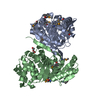

 Mass: 62.068 Da / Num. of mol.: 3 / Source method: obtained synthetically / Formula: C2H6O2
 Mass: 106.120 Da / Num. of mol.: 1 / Source method: obtained synthetically / Formula: C4H10O3
 Mass: 18.015 Da / Num. of mol.: 397 / Source method: isolated from a natural source / Formula: H2O
 Sample preparation
 / Beamline: BL11-1 / Wavelength: 0.97775
  Processing