 Movie
Movie Controller

[English] 日本語
 Yorodumi
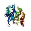
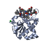
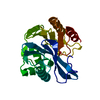
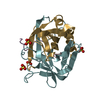
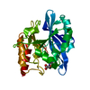
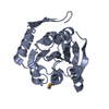
- PDB-2p14: Crystal structure of small subunit (R.BspD6I2) of the heterodimer... -
+ Open data

- Basic information
| Entry | Database: PDB / ID: 2p14 | ||||||
|---|---|---|---|---|---|---|---|
| Title | Crystal structure of small subunit (R.BspD6I2) of the heterodimeric restriction endonuclease R.BspD6I | ||||||
 Components Components | Heterodimeric restriction endonuclease R.BspD6I small subunit | ||||||
 Keywords Keywords | HYDROLASE / heterodimeric restriction endonuclease | ||||||
| Function / homology | Restriction Endonuclease - #50 / Restriction endonuclease, type II, AlwI / AlwI restriction endonuclease / Restriction Endonuclease / endonuclease activity / 3-Layer(aba) Sandwich / Alpha Beta / Heterodimeric restriction endonuclease R.BspD6I small subunit Function and homology information Function and homology information | ||||||
| Biological species |  | ||||||
| Method |  X-RAY DIFFRACTION / SYNCHROTRON / MOLECULAR REPLACEMENT / Resolution: 1.5 Å X-RAY DIFFRACTION / SYNCHROTRON / MOLECULAR REPLACEMENT / Resolution: 1.5 Å | ||||||
 Authors Authors | Kachalova, G.S. / Bartunik, H.D. / Artyukh, R.I. / Rogulin, E.A. / Yunusova, A.K. / Zheleznaya, L.A. / Matvienko, N.I. | ||||||
 Citation Citation | Journal: J.Mol.Biol. / Year: 2008 Title: Structural analysis of the heterodimeric type IIS restriction endonuclease R.BspD6I acting as a complex between a monomeric site-specific nickase and a catalytic subunit. Authors: Kachalova, G.S. / Rogulin, E.A. / Yunusova, A.K. / Artyukh, R.I. / Perevyazova, T.A. / Matvienko, N.I. / Zheleznaya, L.A. / Bartunik, H.D. | ||||||
| History |
|
- Structure visualization
| Structure viewer | Molecule: MolmilJmol/JSmol |
|---|
- Downloads & links
-Download
| PDBx/mmCIF format | 2p14.cif.gz | 56.7 KB | Display | PDBx/mmCIF format |
|---|---|---|---|---|
| PDB format | pdb2p14.ent.gz | 40.2 KB | Display | PDB format |
| PDBx/mmJSON format | 2p14.json.gz | Tree view | PDBx/mmJSON format | |
| Others |  Other downloads Other downloads |
-Validation report
| Arichive directory | https://data.pdbj.org/pub/pdb/validation_reports/p1/2p14ftp://data.pdbj.org/pub/pdb/validation_reports/p1/2p14 | HTTPS FTP |
|---|
-Related structure data
| Related structure data | 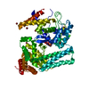 2ewfSC S: Starting model for refinement C: citing same article ( |
|---|---|
| Similar structure data |


-Links
 PDBj
PDBj- Assembly
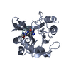
| Deposited unit | 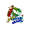
| ||||||||
|---|---|---|---|---|---|---|---|---|---|
| 1 |
| ||||||||
| Unit cell |
|
-Components
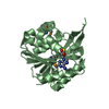
| #1: Protein | Mass: 22508.512 Da / Num. of mol.: 1 Source method: isolated from a genetically manipulated source Source: (gene. exp.) References: UniProt: A3FEV8, type II site-specific deoxyribonuclease | ||||
|---|---|---|---|---|---|
| #2: Chemical |   Mass: 96.063 Da / Num. of mol.: 2 / Source method: obtained synthetically / Formula: SO4 Mass: 96.063 Da / Num. of mol.: 2 / Source method: obtained synthetically / Formula: SO4#3: Chemical | ChemComp-GOL / |   Mass: 92.094 Da / Num. of mol.: 1 / Source method: obtained synthetically / Formula: C3H8O3 Mass: 92.094 Da / Num. of mol.: 1 / Source method: obtained synthetically / Formula: C3H8O3#4: Water | ChemComp-HOH / |  Mass: 18.015 Da / Num. of mol.: 159 / Source method: isolated from a natural source / Formula: H2O Mass: 18.015 Da / Num. of mol.: 159 / Source method: isolated from a natural source / Formula: H2O |
-Experimental details
-Experiment
| Experiment | Method: X-RAY DIFFRACTION / Number of used crystals: 1 |
|---|
- Sample preparation
| Crystal | Density Matthews: 1.99 Å3/Da / Density % sol: 38.18 % |
|---|---|
| Crystal grow | Temperature: 298 K / Method: vapor diffusion, sitting drop / pH: 10.5 Details: 2.0 M (NH4)2SO4, 0.2 M Li2SO4, pH 10.5, VAPOR DIFFUSION, SITTING DROP, temperature 298.0K |
-Data collection
| Diffraction | Mean temperature: 100 K |
|---|---|
| Diffraction source | Source: SYNCHROTRON / Site: MPG/DESY, HAMBURG  / Beamline: BW6 / Wavelength: 1.05 Å / Beamline: BW6 / Wavelength: 1.05 Å |
| Detector | Type: MAR CCD 165 mm / Detector: CCD / Date: Dec 17, 2006 / Details: AU-COATED PLANAR and TOROIDAL MIRRORS |
| Radiation | Monochromator: SI111 / Protocol: SINGLE WAVELENGTH / Monochromatic (M) / Laue (L): M / Scattering type: x-ray |
| Radiation wavelength | Wavelength: 1.05 Å / Relative weight: 1 |
| Reflection | Resolution: 1.5→20 Å / Num. all: 29298 / Num. obs: 28430 / % possible obs: 99.3 % / Observed criterion σ(I): 2 / Redundancy: 2.91 % / Biso Wilson estimate: 18.66 Å2 / Rmerge(I) obs: 0.054 / Net I/σ(I): 21.2 |
| Reflection shell | Resolution: 1.5→1.53 Å / Redundancy: 2.87 % / Rmerge(I) obs: 0.47 / Mean I/σ(I) obs: 2.24 / Num. unique all: 1428 |
- Processing
| Software |
| ||||||||||||||||||||||||||||||||||||||||||||||||||||||||||||||||||||||||||||||||||||||||||
|---|---|---|---|---|---|---|---|---|---|---|---|---|---|---|---|---|---|---|---|---|---|---|---|---|---|---|---|---|---|---|---|---|---|---|---|---|---|---|---|---|---|---|---|---|---|---|---|---|---|---|---|---|---|---|---|---|---|---|---|---|---|---|---|---|---|---|---|---|---|---|---|---|---|---|---|---|---|---|---|---|---|---|---|---|---|---|---|---|---|---|---|
| Refinement | Method to determine structure: MOLECULAR REPLACEMENT Starting model: PDB ENTRY 2ewf Resolution: 1.5→10.75 Å / Cor.coef. Fo:Fc: 0.965 / Cor.coef. Fo:Fc free: 0.955 / SU B: 1.399 / SU ML: 0.053 / Cross valid method: THROUGHOUT / σ(F): 0 / ESU R: 0.081 / ESU R Free: 0.083 / Stereochemistry target values: MAXIMUM LIKELIHOOD
| ||||||||||||||||||||||||||||||||||||||||||||||||||||||||||||||||||||||||||||||||||||||||||
| Solvent computation | Ion probe radii: 0.8 Å / Shrinkage radii: 0.8 Å / VDW probe radii: 1.2 Å / Solvent model: MASK | ||||||||||||||||||||||||||||||||||||||||||||||||||||||||||||||||||||||||||||||||||||||||||
| Displacement parameters | Biso mean: 18.891 Å2
| ||||||||||||||||||||||||||||||||||||||||||||||||||||||||||||||||||||||||||||||||||||||||||
| Refinement step | Cycle: LAST / Resolution: 1.5→10.75 Å
| ||||||||||||||||||||||||||||||||||||||||||||||||||||||||||||||||||||||||||||||||||||||||||
| Refine LS restraints |
| ||||||||||||||||||||||||||||||||||||||||||||||||||||||||||||||||||||||||||||||||||||||||||
| LS refinement shell | Resolution: 1.5→1.538 Å / Total num. of bins used: 20
|
