 Movie
Movie Controller

[English] 日本語
 Yorodumi
- PDB-1p3m: Crystallographic Studies of Nucleosome Core Particles containing ... -
+ Open data

- Basic information
| Entry | Database: PDB / ID: 1p3m | ||||||
|---|---|---|---|---|---|---|---|
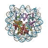
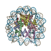
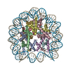
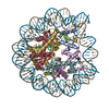
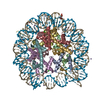
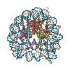
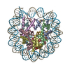
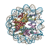
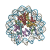
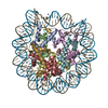
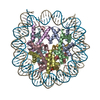
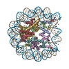
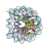
| Title | Crystallographic Studies of Nucleosome Core Particles containing Histone 'Sin' Mutants | ||||||
 Components Components |
| ||||||
 Keywords Keywords | STRUCTURAL PROTEIN/DNA / Sin mutants / Nucleosome Core Particle / chromatin / protein/DNA interaction / STRUCTURAL PROTEIN-DNA COMPLEX | ||||||
| Function / homology |  Function and homology information Function and homology informationstructural constituent of chromatin / nucleosome / heterochromatin formation / nucleosome assembly / protein heterodimerization activity / DNA binding / nucleoplasm / nucleus Similarity search - Function | ||||||
| Biological species |  Homo sapiens (human) Homo sapiens (human) | ||||||
| Method |  X-RAY DIFFRACTION / MOLECULAR REPLACEMENT / Resolution: 2.9 Å X-RAY DIFFRACTION / MOLECULAR REPLACEMENT / Resolution: 2.9 Å | ||||||
 Authors Authors | Muthurajan, U.M. / Bao, Y. / Forsberg, L.J. / Edayathumangalam, R.S. / Dyer, P.N. / White, C.L. / Luger, K. | ||||||
 Citation Citation | Journal: EMBO J. / Year: 2004 Title: Crystal structures of histone Sin mutant nucleosomes reveal altered protein-DNA interactions Authors: Muthurajan, U.M. / Bao, Y. / Forsberg, L.J. / Edayathumangalam, R.S. / Dyer, P.N. / White, C.L. / Luger, K. | ||||||
| History |
|
- Structure visualization
| Structure viewer | Molecule: MolmilJmol/JSmol |
|---|
- Downloads & links
-Download
| PDBx/mmCIF format | 1p3m.cif.gz | 324.5 KB | Display | PDBx/mmCIF format |
|---|---|---|---|---|
| PDB format | pdb1p3m.ent.gz | 246.5 KB | Display | PDB format |
| PDBx/mmJSON format | 1p3m.json.gz | Tree view | PDBx/mmJSON format | |
| Others |  Other downloads Other downloads |
-Validation report
| Arichive directory | https://data.pdbj.org/pub/pdb/validation_reports/p3/1p3mftp://data.pdbj.org/pub/pdb/validation_reports/p3/1p3m | HTTPS FTP |
|---|
-Related structure data
| Related structure data | 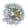 1p34C 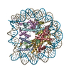 1p3aC 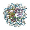 1p3bC 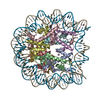 1p3fC  1p3gC  1p3iC  1p3kC  1p3lC  1p3oC  1p3pC  1aoiS S: Starting model for refinement C: citing same article ( |
|---|---|
| Similar structure data |


-Links
 PDBj
PDBj


- Assembly
| Deposited unit | 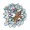
| ||||||||||
|---|---|---|---|---|---|---|---|---|---|---|---|
| 1 |
| ||||||||||
| Unit cell |
|
-Components
-Protein , 4 types, 8 molecules AEBFCGDH
| #2: Protein | Mass: 15375.992 Da / Num. of mol.: 2 Source method: isolated from a genetically manipulated source Source: (gene. exp.)  #3: Protein | Mass: 11263.231 Da / Num. of mol.: 2 Source method: isolated from a genetically manipulated source Source: (gene. exp.) #4: Protein | Mass: 13962.241 Da / Num. of mol.: 2 Source method: isolated from a genetically manipulated source Source: (gene. exp.) #5: Protein | Mass: 13834.070 Da / Num. of mol.: 2 Source method: isolated from a genetically manipulated source Source: (gene. exp.) |
|---|
-DNA chain / Non-polymers , 2 types, 119 molecules IJ
| #1: DNA chain | Mass: 45054.844 Da / Num. of mol.: 2 Source method: isolated from a genetically manipulated source Source: (gene. exp.) Homo sapiens (human) / Plasmid: pUC / Production host: #6: Water | ChemComp-HOH / | Mass: 18.015 Da / Num. of mol.: 117 / Source method: isolated from a natural source / Formula: H2O |
|---|
-Experimental details
-Experiment
| Experiment | Method: X-RAY DIFFRACTION / Number of used crystals: 1 |
|---|
- Sample preparation
| Crystal | Density Matthews: 2.6 Å3/Da / Density % sol: 52.71 % | ||||||||||||||||||||||||||||||||||||||||||||||||||||||||
|---|---|---|---|---|---|---|---|---|---|---|---|---|---|---|---|---|---|---|---|---|---|---|---|---|---|---|---|---|---|---|---|---|---|---|---|---|---|---|---|---|---|---|---|---|---|---|---|---|---|---|---|---|---|---|---|---|---|
| Crystal grow | Temperature: 292 K / Method: vapor diffusion, sitting drop / pH: 6 Details: MnCl2, KCl, Potassium cacodylate, pH 6.0, VAPOR DIFFUSION, SITTING DROP, temperature 292K | ||||||||||||||||||||||||||||||||||||||||||||||||||||||||
| Components of the solutions |
| ||||||||||||||||||||||||||||||||||||||||||||||||||||||||
| Crystal grow | *PLUS Temperature: 20 ℃ / Method: vapor diffusion / Details: Luger, K., (1997) Nature, 389, 251. | ||||||||||||||||||||||||||||||||||||||||||||||||||||||||
| Components of the solutions | *PLUS
|
-Data collection
| Diffraction | Mean temperature: 100 K |
|---|---|
| Diffraction source | Source: ROTATING ANODE / Type: RIGAKU / Wavelength: 1.5418 Å |
| Detector | Type: RIGAKU RAXIS IV / Detector: IMAGE PLATE / Date: Mar 15, 2002 / Details: Mirrors |
| Radiation | Monochromator: Cu / Protocol: SINGLE WAVELENGTH / Monochromatic (M) / Laue (L): M / Scattering type: x-ray |
| Radiation wavelength | Wavelength: 1.5418 Å / Relative weight: 1 |
| Reflection | Resolution: 2.9→35 Å / Num. obs: 37684 / % possible obs: 87.6 % / Observed criterion σ(I): -3 / Redundancy: 1.67 % / Rmerge(I) obs: 0.071 / Net I/σ(I): 20.9 |
| Reflection shell | Resolution: 2.9→2.97 Å / Rmerge(I) obs: 0.377 / Mean I/σ(I) obs: 2.3 / % possible all: 84.8 |
| Reflection | *PLUS Highest resolution: 2.9 Å / Num. obs: 43053 / % possible obs: 87.4 % |
| Reflection shell | *PLUS % possible obs: 84.8 % |
- Processing
| Software |
| |||||||||||||||||||||||||
|---|---|---|---|---|---|---|---|---|---|---|---|---|---|---|---|---|---|---|---|---|---|---|---|---|---|---|
| Refinement | Method to determine structure: MOLECULAR REPLACEMENT Starting model: PDB entry 1AOI Resolution: 2.9→35 Å / Cross valid method: THROUGHOUT / σ(F): 0 / σ(I): 0 / Stereochemistry target values: Engh & Huber
| |||||||||||||||||||||||||
| Refinement step | Cycle: LAST / Resolution: 2.9→35 Å
| |||||||||||||||||||||||||
| Refine LS restraints |
| |||||||||||||||||||||||||
| Refinement | *PLUS Highest resolution: 2.9 Å / Rfactor Rwork: 0.21 | |||||||||||||||||||||||||
| Solvent computation | *PLUS | |||||||||||||||||||||||||
| Displacement parameters | *PLUS | |||||||||||||||||||||||||
| Refine LS restraints | *PLUS Type: c_angle_d | |||||||||||||||||||||||||
| LS refinement shell | *PLUS Rfactor Rfree: 0.42 / Rfactor Rwork: 0.38 |
