 Movie
Movie Controller


+ Open data

- Basic information
| Entry | Database: PDB / ID: 1h1v | |||||||||
|---|---|---|---|---|---|---|---|---|---|---|
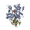
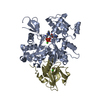
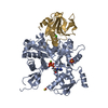
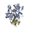
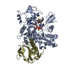
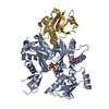
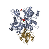
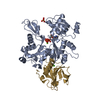
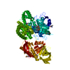
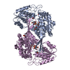
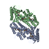
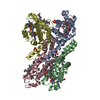
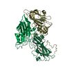
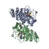
| Title | gelsolin G4-G6/actin complex | |||||||||
 Components Components |
| |||||||||
 Keywords Keywords | ACTIN-BINDING / SEVERING / CAPPING / CALCIUM / AMYLOID / MUSCLE CONTRACTION | |||||||||
| Function / homology |  Function and homology information Function and homology informationStriated Muscle Contraction / dynactin complex / striated muscle atrophy / regulation of establishment of T cell polarity / regulation of plasma membrane raft polarization / regulation of receptor clustering / positive regulation of keratinocyte apoptotic process / renal protein absorption / positive regulation of protein processing in phagocytic vesicle / phosphatidylinositol 3-kinase catalytic subunit binding ...Striated Muscle Contraction / dynactin complex / striated muscle atrophy / regulation of establishment of T cell polarity / regulation of plasma membrane raft polarization / regulation of receptor clustering / positive regulation of keratinocyte apoptotic process / renal protein absorption / positive regulation of protein processing in phagocytic vesicle / phosphatidylinositol 3-kinase catalytic subunit binding / positive regulation of actin nucleation / actin cap / myosin II binding / host-mediated suppression of symbiont invasion / cell projection assembly / actin filament severing / barbed-end actin filament capping / actin polymerization or depolymerization / actin filament depolymerization / actin filament capping / relaxation of cardiac muscle / Sensory processing of sound by outer hair cells of the cochlea / phagocytosis, engulfment / cardiac muscle cell contraction / hepatocyte apoptotic process / cytoskeletal motor activator activity / myosin heavy chain binding / tropomyosin binding / actin filament bundle / troponin I binding / filamentous actin / mesenchyme migration / skeletal muscle myofibril / sarcoplasm / cilium assembly / actin filament bundle assembly / striated muscle thin filament / skeletal muscle thin filament assembly / actin monomer binding / Caspase-mediated cleavage of cytoskeletal proteins / skeletal muscle fiber development / phagocytic vesicle / response to muscle stretch / stress fiber / titin binding / actin filament polymerization / phosphatidylinositol-4,5-bisphosphate binding / actin filament organization / central nervous system development / filopodium / actin filament / cellular response to type II interferon / protein destabilization / Hydrolases; Acting on acid anhydrides; Acting on acid anhydrides to facilitate cellular and subcellular movement / calcium-dependent protein binding / actin filament binding / lamellipodium / actin cytoskeleton / actin binding / cell body / secretory granule lumen / blood microparticle / amyloid fibril formation / ficolin-1-rich granule lumen / Amyloid fiber formation / protein domain specific binding / focal adhesion / hydrolase activity / calcium ion binding / positive regulation of gene expression / Neutrophil degranulation / magnesium ion binding / : / extracellular exosome / extracellular region / ATP binding / identical protein binding / plasma membrane / cytosol / cytoplasm Similarity search - Function | |||||||||
| Biological species |  HOMO SAPIENS (human) HOMO SAPIENS (human) | |||||||||
| Method |  X-RAY DIFFRACTION / SYNCHROTRON / MAD / Resolution: 2.99 Å X-RAY DIFFRACTION / SYNCHROTRON / MAD / Resolution: 2.99 Å | |||||||||
 Authors Authors | Choe, H. / Burtnick, L.D. / Mejillano, M. / Yin, H.L. / Robinson, R.C. / Choe, S. | |||||||||
 Citation Citation | Journal: J.Mol.Biol. / Year: 2002 Title: The Calcium Activation of Gelsolin:Insights from the 3A Structure of the G4-G6/Actin Complex Authors: Choe, H. / Burtnick, L.D. / Mejillano, M. / Yin, H.L. / Robinson, R.C. / Choe, S. #1: Journal: Science / Year: 1999 Title: Domain Movement in Gelsolin: A Calcium-Activated Switch Authors: Choe, H. / Burtnick, L.D. / Mejillano, M. / Yin, H.L. / Robinson, R.C. / Choe, S. | |||||||||
| History |
| |||||||||
| Remark 700 | SHEET THE SHEET STRUCTURE OF THIS MOLECULE IS BIFURCATED. IN ORDER TO REPRESENT THIS FEATURE IN ... SHEET THE SHEET STRUCTURE OF THIS MOLECULE IS BIFURCATED. IN ORDER TO REPRESENT THIS FEATURE IN THE SHEET RECORDS BELOW, TWO SHEETS ARE DEFINED. |
- Structure visualization
| Structure viewer | Molecule: MolmilJmol/JSmol |
|---|
- Downloads & links
-Download
| PDBx/mmCIF format | 1h1v.cif.gz | 161.2 KB | Display | PDBx/mmCIF format |
|---|---|---|---|---|
| PDB format | pdb1h1v.ent.gz | 123.3 KB | Display | PDB format |
| PDBx/mmJSON format | 1h1v.json.gz | Tree view | PDBx/mmJSON format | |
| Others |  Other downloads Other downloads |
-Validation report
| Arichive directory | https://data.pdbj.org/pub/pdb/validation_reports/h1/1h1vftp://data.pdbj.org/pub/pdb/validation_reports/h1/1h1v | HTTPS FTP |
|---|
-Related structure data
| Related structure data |  1db0 S: Starting model for refinement |
|---|---|
| Similar structure data |


-Links
 PDBj
PDBj


- Assembly
| Deposited unit | 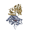
| ||||||||
|---|---|---|---|---|---|---|---|---|---|
| 1 |
| ||||||||
| Unit cell |
| ||||||||
| Details | THE COMPLEX IS A HETERODIMER WITH ONE MOLECULE OF ACTINAND ONE MOLECULE OF GELSOLIN. |
-Components
| #1: Protein | Mass: 41862.613 Da / Num. of mol.: 1 / Source method: isolated from a natural source / Source: (natural) | ||||||||
|---|---|---|---|---|---|---|---|---|---|
| #2: Protein | Mass: 36407.551 Da / Num. of mol.: 1 / Fragment: G4-G6, RESIDUES 412-742 OF CYTOPLASMIC ISOFORM Source method: isolated from a genetically manipulated source Source: (gene. exp.) HOMO SAPIENS (human) / Production host:  | ||||||||
| #3: Chemical | ChemComp-CA /   Mass: 40.078 Da / Num. of mol.: 5 / Source method: obtained synthetically / Formula: Ca Mass: 40.078 Da / Num. of mol.: 5 / Source method: obtained synthetically / Formula: Ca#4: Chemical | ChemComp-ATP / |   Mass: 507.181 Da / Num. of mol.: 1 / Source method: obtained synthetically / Formula: C10H16N5O13P3 / Comment: ATP, energy-carrying molecule*YM Mass: 507.181 Da / Num. of mol.: 1 / Source method: obtained synthetically / Formula: C10H16N5O13P3 / Comment: ATP, energy-carrying molecule*YM#5: Water | ChemComp-HOH / |  Mass: 18.015 Da / Num. of mol.: 346 / Source method: isolated from a natural source / Formula: H2O Mass: 18.015 Da / Num. of mol.: 346 / Source method: isolated from a natural source / Formula: H2OCompound details | GELSOLIN: CALCIUM-REGULATED, ACTIN-MODULATING PROTEIN THAT BINDS TO ACTIN MONOMERS OR FILAMENTS AT ...GELSOLIN: CALCIUM-REGULATED, ACTIN-MODULATING | Sequence details | SWISSPROT HAS MERGED THE SEQUENCE OF RABBIT, HUMAN PIG, RAT AND MOUSE ACTIN AND GIVEN THE SWISSPROT ID P02568. | |
-Experimental details
-Experiment
| Experiment | Method: X-RAY DIFFRACTION / Number of used crystals: 1 |
|---|
- Sample preparation
| Crystal | Density Matthews: 3.14 Å3/Da / Density % sol: 60.81 % | |||||||||||||||||||||||||||||||||||||||||||||||||||||||||||||||
|---|---|---|---|---|---|---|---|---|---|---|---|---|---|---|---|---|---|---|---|---|---|---|---|---|---|---|---|---|---|---|---|---|---|---|---|---|---|---|---|---|---|---|---|---|---|---|---|---|---|---|---|---|---|---|---|---|---|---|---|---|---|---|---|---|
| Crystal grow | pH: 7.5 Details: PROTEIN WAS CRYSTALLIZED FROM 100MM HEPES BUFFER, PH 7.5, 20% GLYCEROL, 10 % PEG 8000 | |||||||||||||||||||||||||||||||||||||||||||||||||||||||||||||||
| Crystal grow | *PLUS Temperature: 4 ℃ / pH: 8 / Method: vapor diffusion / Details: Robinson, R.C., (1999) Science, 286, 1939. | |||||||||||||||||||||||||||||||||||||||||||||||||||||||||||||||
| Components of the solutions | *PLUS
|
-Data collection
| Diffraction | Mean temperature: 95 K |
|---|---|
| Diffraction source | Source: SYNCHROTRON / Site: SSRL  / Beamline: BL9-2 / Wavelength: 1.07 / Beamline: BL9-2 / Wavelength: 1.07 |
| Detector | Detector: CCD |
| Radiation | Protocol: SINGLE WAVELENGTH / Monochromatic (M) / Laue (L): M / Scattering type: x-ray |
| Radiation wavelength | Wavelength: 1.07 Å / Relative weight: 1 |
| Reflection | Resolution: 3→20 Å / Num. obs: 20076 / % possible obs: 98 % / Observed criterion σ(I): 0 / Redundancy: 4.3 % / Rmerge(I) obs: 0.074 / Net I/σ(I): 8.2 |
| Reflection shell | Rmerge(I) obs: 0.3 |
| Reflection | *PLUS Lowest resolution: 20 Å / % possible obs: 98 % |
- Processing
| Software |
| ||||||||||||||||||||
|---|---|---|---|---|---|---|---|---|---|---|---|---|---|---|---|---|---|---|---|---|---|
| Refinement | Method to determine structure: MAD Starting model: PDB ENTRY 1DB0 1db0 Resolution: 2.99→19.73 Å / SU B: 24.358 / SU ML: 0.464 / Cross valid method: THROUGHOUT / ESU R Free: 0.45
| ||||||||||||||||||||
| Displacement parameters | Biso mean: 26.173 Å2
| ||||||||||||||||||||
| Refinement step | Cycle: LAST / Resolution: 2.99→19.73 Å
| ||||||||||||||||||||
| Refinement | *PLUS Highest resolution: 3 Å / Lowest resolution: 20 Å | ||||||||||||||||||||
| Solvent computation | *PLUS | ||||||||||||||||||||
| Displacement parameters | *PLUS | ||||||||||||||||||||
| Refine LS restraints | *PLUS
| ||||||||||||||||||||
| LS refinement shell | *PLUS Highest resolution: 2.994 Å / Lowest resolution: 3.07 Å / Rfactor Rfree: 0.414 / % reflection Rfree: 65 % / Rfactor Rwork: 0.295 / Num. reflection Rwork: 1141 / Total num. of bins used: 20 |
