 Movie
Movie Controller


+ Open data

- Basic information
| Entry | Database: PDB / ID: 3zx3 | ||||||
|---|---|---|---|---|---|---|---|
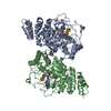
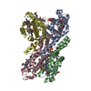
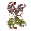
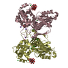
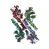
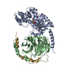
| Title | Crystal Structure and Domain Rotation of NTPDase1 CD39 | ||||||
 Components Components | ECTONUCLEOSIDE TRIPHOSPHATE DIPHOSPHOHYDROLASE 1 | ||||||
 Keywords Keywords | HYDROLASE / DOMAIN ROTATION / PURINERGIC SIGNALING | ||||||
| Function / homology |  Function and homology information Function and homology informationresponse to proline / CDP phosphatase activity / ITPase activity / purine ribonucleoside diphosphate catabolic process / Phosphate bond hydrolysis by NTPDase proteins / apyrase activity / apyrase / UDP phosphatase activity / IDP phosphatase activity / CTPase activity ...response to proline / CDP phosphatase activity / ITPase activity / purine ribonucleoside diphosphate catabolic process / Phosphate bond hydrolysis by NTPDase proteins / apyrase activity / apyrase / UDP phosphatase activity / IDP phosphatase activity / CTPase activity / GDP phosphatase activity / ADP phosphatase activity / nucleoside diphosphate catabolic process / cellular response to interferon-alpha / cellular response to aluminum ion / ADP catabolic process / nucleoside diphosphate phosphatase activity / negative regulation of dopamine secretion / response to L-arginine / negative regulation of ATP biosynthetic process / response to auditory stimulus / response to caffeine / response to ATP / basement membrane / response to gamma radiation / synaptic membrane / platelet activation / caveola / platelet aggregation / cellular response to tumor necrosis factor / ribonucleoside triphosphate phosphatase activity / synaptic vesicle / cellular response to lipopolysaccharide / response to lipopolysaccharide / basolateral plasma membrane / G protein-coupled receptor signaling pathway / external side of plasma membrane / neuronal cell body / GTPase activity / GTP binding / cell surface / ATP hydrolysis activity / : / ATP binding / identical protein binding / plasma membrane Similarity search - Function | ||||||
| Biological species |  | ||||||
| Method |  X-RAY DIFFRACTION / SYNCHROTRON / MOLECULAR REPLACEMENT / Resolution: 1.7 Å X-RAY DIFFRACTION / SYNCHROTRON / MOLECULAR REPLACEMENT / Resolution: 1.7 Å | ||||||
 Authors Authors | Zebisch, M. / Schaefer, P. / Straeter, N. | ||||||
 Citation Citation | Journal: J.Mol.Biol. / Year: 2012 Title: Crystallographic Evidence for a Domain Motion in Rat Nucleoside Triphosphate Diphosphohydrolase (Ntpdase) 1. Authors: Zebisch, M. / Krauss, M. / Schafer, P. / Strater, N. | ||||||
| History |
|
- Structure visualization
| Structure viewer | Molecule: MolmilJmol/JSmol |
|---|
- Downloads & links
-Download
| PDBx/mmCIF format | 3zx3.cif.gz | 658.5 KB | Display | PDBx/mmCIF format |
|---|---|---|---|---|
| PDB format | pdb3zx3.ent.gz | 544.6 KB | Display | PDB format |
| PDBx/mmJSON format | 3zx3.json.gz | Tree view | PDBx/mmJSON format | |
| Others |  Other downloads Other downloads |
-Validation report
| Arichive directory | https://data.pdbj.org/pub/pdb/validation_reports/zx/3zx3ftp://data.pdbj.org/pub/pdb/validation_reports/zx/3zx3 | HTTPS FTP |
|---|
-Related structure data
| Related structure data | 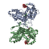 3zx0C  3zx2C  3cj1S C: citing same article ( S: Starting model for refinement |
|---|---|
| Similar structure data |


-Links
 PDBj
PDBj

- Assembly
| Deposited unit | 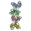
| ||||||||
|---|---|---|---|---|---|---|---|---|---|
| 1 | 
| ||||||||
| 2 | 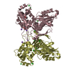
| ||||||||
| Unit cell |
|
-Components
| #1: Protein | Mass: 50576.953 Da / Num. of mol.: 4 / Fragment: ECTODOMAIN, RESIDUES 38-189,190-206 Source method: isolated from a genetically manipulated source Details: A PUTATIVE MEMBRANE INTERACTION LOOP 190TQEQSWLNFISDSQKQA206 WAS REPLACED BY A SHORTER LINKER KTPGGS Source: (gene. exp.)  #2: Chemical | ChemComp-CL /   Mass: 35.453 Da / Num. of mol.: 27 / Source method: obtained synthetically / Formula: Cl Mass: 35.453 Da / Num. of mol.: 27 / Source method: obtained synthetically / Formula: Cl#3: Chemical | ChemComp-ACY /   Mass: 60.052 Da / Num. of mol.: 7 / Source method: obtained synthetically / Formula: C2H4O2 Mass: 60.052 Da / Num. of mol.: 7 / Source method: obtained synthetically / Formula: C2H4O2#4: Chemical | ChemComp-NA /   Mass: 22.990 Da / Num. of mol.: 5 / Source method: obtained synthetically / Formula: Na Mass: 22.990 Da / Num. of mol.: 5 / Source method: obtained synthetically / Formula: Na#5: Water | ChemComp-HOH / |  Mass: 18.015 Da / Num. of mol.: 816 / Source method: isolated from a natural source / Formula: H2O Mass: 18.015 Da / Num. of mol.: 816 / Source method: isolated from a natural source / Formula: H2OHas protein modification | Y | Sequence details | THE LOOP T190 TO A206 WAS REPLACED WITH THE SEQUENCE KTPGGS OF GB EDL94186.1. THE RESIDUES 80, 220, ...THE LOOP T190 TO A206 WAS REPLACED WITH THE SEQUENCE KTPGGS OF GB EDL94186.1. THE RESIDUES 80, 220, AND 227 MATCH THE GENBANK SEQUENCE. | |
|---|
-Experimental details
-Experiment
| Experiment | Method: X-RAY DIFFRACTION / Number of used crystals: 1 |
|---|
- Sample preparation
| Crystal | Density Matthews: 2.4 Å3/Da / Density % sol: 49 % / Description: NONE |
|---|---|
| Crystal grow | pH: 4.5 / Details: 3.6 M NACL, 100 MM NAAC PH 4.5. |
-Data collection
| Diffraction | Mean temperature: 100 K |
|---|---|
| Diffraction source | Source: SYNCHROTRON / Site: BESSY  / Beamline: 14.2 / Wavelength: 0.9184 / Beamline: 14.2 / Wavelength: 0.9184 |
| Detector | Type: MARMOSAIC 225 mm CCD / Detector: CCD / Date: Sep 9, 2009 |
| Radiation | Protocol: SINGLE WAVELENGTH / Monochromatic (M) / Laue (L): M / Scattering type: x-ray |
| Radiation wavelength | Wavelength: 0.9184 Å / Relative weight: 1 |
| Reflection | Resolution: 1.7→42.55 Å / Num. obs: 151721 / % possible obs: 72.4 % / Observed criterion σ(I): -3 / Redundancy: 5.6 % / Rmerge(I) obs: 0.06 / Net I/σ(I): 13.8 |
| Reflection shell | Resolution: 1.7→1.8 Å / Redundancy: 4.9 % / Rmerge(I) obs: 0.54 / Mean I/σ(I) obs: 2.4 / % possible all: 7.7 |
- Processing
| Software |
| ||||||||||||||||||||
|---|---|---|---|---|---|---|---|---|---|---|---|---|---|---|---|---|---|---|---|---|---|
| Refinement | Method to determine structure: MOLECULAR REPLACEMENT Starting model: PDB ENTRY 3CJ1 Resolution: 1.7→146.62 Å / Cor.coef. Fo:Fc: 0.972 / Cor.coef. Fo:Fc free: 0.948 / Cross valid method: THROUGHOUT / ESU R: 0.234 / ESU R Free: 0.133 / Stereochemistry target values: MAXIMUM LIKELIHOOD / Details: HYDROGENS HAVE BEEN USED IF PRESENT IN THE INPUT
| ||||||||||||||||||||
| Solvent computation | Ion probe radii: 0.8 Å / Shrinkage radii: 0.8 Å / VDW probe radii: 1.2 Å / Solvent model: MASK | ||||||||||||||||||||
| Displacement parameters | Biso mean: 45.504 Å2
| ||||||||||||||||||||
| Refinement step | Cycle: LAST / Resolution: 1.7→146.62 Å
| ||||||||||||||||||||
| LS refinement shell | Resolution: 1.703→1.747 Å / Total num. of bins used: 20
|
