 Movie
Movie Controller

[English] 日本語
 Yorodumi
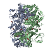
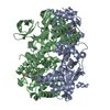
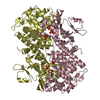
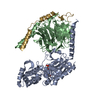
- PDB-3ah8: Structure of heterotrimeric G protein Galpha-q beta gamma in comp... -
+ Open data

- Basic information
| Entry | Database: PDB / ID: 3ah8 | |||||||||
|---|---|---|---|---|---|---|---|---|---|---|
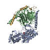
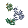
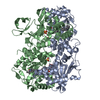
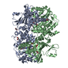
| Title | Structure of heterotrimeric G protein Galpha-q beta gamma in complex with an inhibitor YM-254890 | |||||||||
 Components Components |
| |||||||||
 Keywords Keywords | SIGNALING PROTEIN/INHIBITOR / Heterotrimeric G protein / GTPase / Galpha-q / Gbeta / Ggamma / Inhibitor / YM-254890 / SIGNALING PROTEIN / SIGNALING PROTEIN-INHIBITOR complex | |||||||||
| Function / homology |  Function and homology information Function and homology informationFatty Acids bound to GPR40 (FFAR1) regulate insulin secretion / PLC beta mediated events / Acetylcholine regulates insulin secretion / forebrain neuron development / regulation of melanocyte differentiation / Cooperation of PDCL (PhLP1) and TRiC/CCT in G-protein beta folding / Thromboxane signalling through TP receptor / adenylate cyclase regulator activity / Extra-nuclear estrogen signaling / Thrombin signalling through proteinase activated receptors (PARs) ...Fatty Acids bound to GPR40 (FFAR1) regulate insulin secretion / PLC beta mediated events / Acetylcholine regulates insulin secretion / forebrain neuron development / regulation of melanocyte differentiation / Cooperation of PDCL (PhLP1) and TRiC/CCT in G-protein beta folding / Thromboxane signalling through TP receptor / adenylate cyclase regulator activity / Extra-nuclear estrogen signaling / Thrombin signalling through proteinase activated receptors (PARs) / adenylate cyclase-activating G protein-coupled cAMP receptor signaling pathway / Adenylate cyclase inhibitory pathway / Turbulent (oscillatory, disturbed) flow shear stress activates signaling by PIEZO1 and integrins in endothelial cells / G-protein activation / G alpha (q) signalling events / phospholipase C-activating G protein-coupled acetylcholine receptor signaling pathway / endothelin receptor signaling pathway / Olfactory Signaling Pathway / Sensory perception of sweet, bitter, and umami (glutamate) taste / Synthesis, secretion, and inactivation of Glucagon-like Peptide-1 (GLP-1) / phospholipase C-activating dopamine receptor signaling pathway / developmental pigmentation / phospholipase C-activating G protein-coupled glutamate receptor signaling pathway / ADP signalling through P2Y purinoceptor 1 / High laminar flow shear stress activates signaling by PIEZO1 and PECAM1:CDH5:KDR in endothelial cells / phospholipase C-activating serotonin receptor signaling pathway / cranial skeletal system development / regulation of platelet activation / Activation of the phototransduction cascade / maternal behavior / negative regulation of synaptic transmission / Adrenaline,noradrenaline inhibits insulin secretion / ADP signalling through P2Y purinoceptor 12 / GTPase activating protein binding / regulation of canonical Wnt signaling pathway / G alpha (i) signalling events / embryonic digit morphogenesis / Activation of G protein gated Potassium channels / G-protein activation / G beta:gamma signalling through PI3Kgamma / Prostacyclin signalling through prostacyclin receptor / G beta:gamma signalling through PLC beta / ADP signalling through P2Y purinoceptor 1 / Thromboxane signalling through TP receptor / Presynaptic function of Kainate receptors / G beta:gamma signalling through CDC42 / Inhibition of voltage gated Ca2+ channels via Gbeta/gamma subunits / G alpha (12/13) signalling events / Glucagon-type ligand receptors / G beta:gamma signalling through BTK / ADP signalling through P2Y purinoceptor 12 / Adrenaline,noradrenaline inhibits insulin secretion / Cooperation of PDCL (PhLP1) and TRiC/CCT in G-protein beta folding / Ca2+ pathway / G alpha (z) signalling events / Thrombin signalling through proteinase activated receptors (PARs) / Extra-nuclear estrogen signaling / G alpha (s) signalling events / G alpha (q) signalling events / glutamate receptor signaling pathway / Glucagon-like Peptide-1 (GLP1) regulates insulin secretion / G alpha (i) signalling events / neuron remodeling / ligand-gated ion channel signaling pathway / neurotransmitter receptor localization to postsynaptic specialization membrane / High laminar flow shear stress activates signaling by PIEZO1 and PECAM1:CDH5:KDR in endothelial cells / Vasopressin regulates renal water homeostasis via Aquaporins / alkylglycerophosphoethanolamine phosphodiesterase activity / negative regulation of potassium ion transport / action potential / negative regulation of insulin secretion / postsynaptic cytosol / hormone-mediated signaling pathway / cellular response to acidic pH / centriolar satellite / enzyme regulator activity / adenylate cyclase inhibitor activity / positive regulation of protein localization to cell cortex / T cell migration / D2 dopamine receptor binding / post-embryonic development / adenylate cyclase-inhibiting serotonin receptor signaling pathway / G protein-coupled serotonin receptor binding / cellular response to forskolin / GTPase activator activity / regulation of mitotic spindle organization / skeletal system development / chemokine-mediated signaling pathway / neuropeptide signaling pathway / response to prostaglandin E / positive regulation of cholesterol biosynthetic process / caveola / G protein-coupled receptor binding / regulation of blood pressure / G-protein beta/gamma-subunit complex binding / adenylate cyclase-modulating G protein-coupled receptor signaling pathway / positive regulation of insulin secretion / adenylate cyclase-inhibiting G protein-coupled receptor signaling pathway / photoreceptor disc membrane / GDP binding Similarity search - Function | |||||||||
| Biological species |   Chromobacterium sp. (bacteria) Chromobacterium sp. (bacteria) | |||||||||
| Method |  X-RAY DIFFRACTION / SYNCHROTRON / MOLECULAR REPLACEMENT / Resolution: 2.9 Å X-RAY DIFFRACTION / SYNCHROTRON / MOLECULAR REPLACEMENT / Resolution: 2.9 Å | |||||||||
 Authors Authors | Nishimura, A. / Kitano, K. / Takasaki, J. / Taniguchi, M. / Mizuno, N. / Tago, K. / Hakoshima, T. / Itoh, H. | |||||||||
 Citation Citation | Journal: Proc.Natl.Acad.Sci.USA / Year: 2010 Title: Structural basis for the specific inhibition of heterotrimeric Gq protein by a small molecule. Authors: Nishimura, A. / Kitano, K. / Takasaki, J. / Taniguchi, M. / Mizuno, N. / Tago, K. / Hakoshima, T. / Itoh, H. | |||||||||
| History |
|
- Structure visualization
| Structure viewer | Molecule: MolmilJmol/JSmol |
|---|
- Downloads & links
-Download
| PDBx/mmCIF format | 3ah8.cif.gz | 159.4 KB | Display | PDBx/mmCIF format |
|---|---|---|---|---|
| PDB format | pdb3ah8.ent.gz | 120.6 KB | Display | PDB format |
| PDBx/mmJSON format | 3ah8.json.gz | Tree view | PDBx/mmJSON format | |
| Others |  Other downloads Other downloads |
-Validation report
| Arichive directory | https://data.pdbj.org/pub/pdb/validation_reports/ah/3ah8ftp://data.pdbj.org/pub/pdb/validation_reports/ah/3ah8 | HTTPS FTP |
|---|
-Related structure data


-Links
 PDBj
PDBj


- Assembly
| Deposited unit | 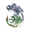
| ||||||||
|---|---|---|---|---|---|---|---|---|---|
| 1 |
| ||||||||
| Unit cell |
|
-Components
| #1: Protein | Mass: 41427.027 Da / Num. of mol.: 1 Fragment: UNP entry P10824 residues 2-28, UNP entry P21279 residues 37-359 Source method: isolated from a genetically manipulated source Source: (gene. exp.) Gene: gmhB / Production host:  Trichoplusia ni (cabbage looper) / Strain (production host): High Five cell / References: UniProt: P10824, UniProt: P21279 Trichoplusia ni (cabbage looper) / Strain (production host): High Five cell / References: UniProt: P10824, UniProt: P21279 |
|---|---|
| #2: Protein | Mass: 37416.930 Da / Num. of mol.: 1 Source method: isolated from a genetically manipulated source Source: (gene. exp.) Trichoplusia ni (cabbage looper) / Strain (production host): High Five cell / References: UniProt: P62871 |
| #3: Protein | Mass: 8592.894 Da / Num. of mol.: 1 / Mutation: C68S Source method: isolated from a genetically manipulated source Source: (gene. exp.) Trichoplusia ni (cabbage looper) / Strain (production host): High Five cell / References: UniProt: P63212 |
| #4: Protein/peptide | 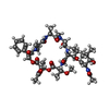  Type: Cyclic depsipeptide / Class: Inhibitor / Mass: 962.094 Da / Num. of mol.: 1 / Source method: isolated from a natural source / Source: (natural) Chromobacterium sp. (bacteria) / Strain: QS3666 / References: YM-254890 Type: Cyclic depsipeptide / Class: Inhibitor / Mass: 962.094 Da / Num. of mol.: 1 / Source method: isolated from a natural source / Source: (natural) Chromobacterium sp. (bacteria) / Strain: QS3666 / References: YM-254890 |
| #5: Chemical | ChemComp-GDP /   Type: RNA linking / Mass: 443.201 Da / Num. of mol.: 1 / Source method: obtained synthetically / Formula: C10H15N5O11P2 / Comment: GDP, energy-carrying molecule*YM Type: RNA linking / Mass: 443.201 Da / Num. of mol.: 1 / Source method: obtained synthetically / Formula: C10H15N5O11P2 / Comment: GDP, energy-carrying molecule*YM |
-Experimental details
-Experiment
| Experiment | Method: X-RAY DIFFRACTION / Number of used crystals: 1 |
|---|
- Sample preparation
| Crystal | Density Matthews: 2.59 Å3/Da / Density % sol: 52.5 % |
|---|---|
| Crystal grow | Temperature: 293 K / Method: vapor diffusion / pH: 5.1 Details: 7% PEG 4000, 30% (v/v) glycerol, 70 mM acetate-NaOH, pH 5.1, VAPOR DIFFUSION, temperature 293K |
-Data collection
| Diffraction | Mean temperature: 100 K |
|---|---|
| Diffraction source | Source: SYNCHROTRON / Site: SPring-8  / Beamline: BL41XU / Wavelength: 1 Å / Beamline: BL41XU / Wavelength: 1 Å |
| Detector | Type: ADSC QUANTUM 315 / Detector: CCD / Date: Nov 24, 2007 |
| Radiation | Protocol: SINGLE WAVELENGTH / Monochromatic (M) / Laue (L): M / Scattering type: x-ray |
| Radiation wavelength | Wavelength: 1 Å / Relative weight: 1 |
| Reflection | Resolution: 2.9→20 Å / Num. obs: 19018 / % possible obs: 94.1 % / Redundancy: 4.2 % / Rmerge(I) obs: 0.059 |
| Reflection shell | Resolution: 2.9→3 Å / Redundancy: 4.1 % / Rmerge(I) obs: 0.526 / Mean I/σ(I) obs: 2.4 / % possible all: 76.9 |
- Processing
| Software |
| ||||||||||||||||||||
|---|---|---|---|---|---|---|---|---|---|---|---|---|---|---|---|---|---|---|---|---|---|
| Refinement | Method to determine structure: MOLECULAR REPLACEMENT Starting model: PDB ENTRIES 2BCJ and 1GP2 Resolution: 2.9→20 Å / Isotropic thermal model: Isotropic / Cross valid method: THROUGHOUT
| ||||||||||||||||||||
| Displacement parameters | Biso mean: 91.53 Å2 | ||||||||||||||||||||
| Refinement step | Cycle: LAST / Resolution: 2.9→20 Å
| ||||||||||||||||||||
| LS refinement shell | Resolution: 2.9→3 Å /
|
