 Movie
Movie Controller

[English] 日本語
 Yorodumi
- PDB-3dkl: Crystal structure of phosphorylated mimic form of human NAMPT com... -
+ Open data

- Basic information
| Entry | Database: PDB / ID: 3dkl | ||||||
|---|---|---|---|---|---|---|---|
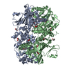
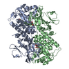
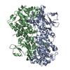
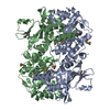
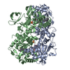
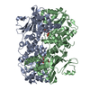
| Title | Crystal structure of phosphorylated mimic form of human NAMPT complexed with benzamide and phosphoribosyl pyrophosphate | ||||||
 Components Components | Nicotinamide phosphoribosyltransferase | ||||||
 Keywords Keywords | TRANSFERASE / NMPRTase / Visfatin / nicotinamide phosphoribosyltransferase / beryllium fluoride / benzamide / phosphoribosyl pyrophosphate / Alternative splicing / Cytoplasm / Glycosyltransferase / Phosphoprotein / Polymorphism / Pyridine nucleotide biosynthesis | ||||||
| Function / homology |  Function and homology information Function and homology informationnicotinamide phosphoribosyltransferase / nicotinamide phosphoribosyltransferase activity / nicotinamide metabolic process / NAD+ biosynthetic process via the salvage pathway / nicotinate metabolic process / NADP+ biosynthetic process / NAD+ biosynthetic process / Nicotinate metabolism / NPAS4 regulates expression of target genes / BMAL1:CLOCK,NPAS2 activates circadian expression ...nicotinamide phosphoribosyltransferase / nicotinamide phosphoribosyltransferase activity / nicotinamide metabolic process / NAD+ biosynthetic process via the salvage pathway / nicotinate metabolic process / NADP+ biosynthetic process / NAD+ biosynthetic process / Nicotinate metabolism / NPAS4 regulates expression of target genes / BMAL1:CLOCK,NPAS2 activates circadian expression / cytokine activity / circadian regulation of gene expression / cell junction / cell-cell signaling / positive regulation of ERK1 and ERK2 cascade / positive regulation of canonical NF-kappaB signal transduction / nuclear speck / mitochondrial matrix / inflammatory response / positive regulation of cell population proliferation / positive regulation of gene expression / signal transduction / positive regulation of transcription by RNA polymerase II / extracellular exosome / identical protein binding / cytosol Similarity search - Function | ||||||
| Biological species |  Homo sapiens (human) Homo sapiens (human) | ||||||
| Method |  X-RAY DIFFRACTION / SYNCHROTRON / FOURIER SYNTHESIS / Resolution: 1.89 Å X-RAY DIFFRACTION / SYNCHROTRON / FOURIER SYNTHESIS / Resolution: 1.89 Å | ||||||
 Authors Authors | Ho, M. / Burgos, E.S. / Almo, S.C. / Schramm, V.L. | ||||||
 Citation Citation | Journal: Proc.Natl.Acad.Sci.USA / Year: 2009 Title: A phosphoenzyme mimic, overlapping catalytic sites and reaction coordinate motion for human NAMPT. Authors: Burgos, E.S. / Ho, M.C. / Almo, S.C. / Schramm, V.L. | ||||||
| History |
|
- Structure visualization
| Structure viewer | Molecule: MolmilJmol/JSmol |
|---|
- Downloads & links
-Download
| PDBx/mmCIF format | 3dkl.cif.gz | 204.9 KB | Display | PDBx/mmCIF format |
|---|---|---|---|---|
| PDB format | pdb3dkl.ent.gz | 162 KB | Display | PDB format |
| PDBx/mmJSON format | 3dkl.json.gz | Tree view | PDBx/mmJSON format | |
| Others |  Other downloads Other downloads |
-Validation report
| Arichive directory | https://data.pdbj.org/pub/pdb/validation_reports/dk/3dklftp://data.pdbj.org/pub/pdb/validation_reports/dk/3dkl | HTTPS FTP |
|---|
-Related structure data
| Related structure data |  3dgrC 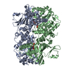 3dhdC 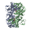 3dhfSC  3dkjC C: citing same article ( S: Starting model for refinement |
|---|---|
| Similar structure data |


-Links
 PDBj
PDBj


- Assembly
| Deposited unit | 
| ||||||||
|---|---|---|---|---|---|---|---|---|---|
| 1 |
| ||||||||
| Unit cell |
|
-Components
-Protein / Sugars , 2 types, 4 molecules AB
| #1: Protein | Mass: 54805.109 Da / Num. of mol.: 2 Source method: isolated from a genetically manipulated source Source: (gene. exp.) Homo sapiens (human) / Gene: NAMPT, PBEF, PBEF1 / Plasmid: pBAD/DEFT 49 / Production host:  References: UniProt: P43490, nicotinamide phosphoribosyltransferase #5: Sugar |  Type: D-saccharide / Mass: 390.070 Da / Num. of mol.: 2 / Source method: obtained synthetically / Formula: C5H13O14P3 Type: D-saccharide / Mass: 390.070 Da / Num. of mol.: 2 / Source method: obtained synthetically / Formula: C5H13O14P3 |
|---|
-Non-polymers , 4 types, 431 molecules 


| #2: Chemical | ChemComp-MG /  Mass: 24.305 Da / Num. of mol.: 4 / Source method: obtained synthetically / Formula: Mg Mass: 24.305 Da / Num. of mol.: 4 / Source method: obtained synthetically / Formula: Mg#3: Chemical |  Mass: 121.137 Da / Num. of mol.: 2 / Source method: obtained synthetically / Formula: C7H7NO Mass: 121.137 Da / Num. of mol.: 2 / Source method: obtained synthetically / Formula: C7H7NO#4: Chemical |  Mass: 66.007 Da / Num. of mol.: 2 / Source method: obtained synthetically / Formula: BeF3 Mass: 66.007 Da / Num. of mol.: 2 / Source method: obtained synthetically / Formula: BeF3#6: Water | ChemComp-HOH / | Mass: 18.015 Da / Num. of mol.: 423 / Source method: isolated from a natural source / Formula: H2O |
|---|
-Experimental details
-Experiment
| Experiment | Method: X-RAY DIFFRACTION / Number of used crystals: 1 |
|---|
- Sample preparation
| Crystal | Density Matthews: 2.46 Å3/Da / Density % sol: 49.93 % |
|---|---|
| Crystal grow | Temperature: 291 K / Method: vapor diffusion, sitting drop / pH: 8.5 Details: 200mM NaCl, 100mM Tris-HCl, 15% PEG 3350, 20% Glycerol, pH 8.5, VAPOR DIFFUSION, SITTING DROP, temperature 291K |
-Data collection
| Diffraction | Mean temperature: 100 K | ||||||||||||||||||||||||||||||||||||||||||||||||||||||||||||||||||
|---|---|---|---|---|---|---|---|---|---|---|---|---|---|---|---|---|---|---|---|---|---|---|---|---|---|---|---|---|---|---|---|---|---|---|---|---|---|---|---|---|---|---|---|---|---|---|---|---|---|---|---|---|---|---|---|---|---|---|---|---|---|---|---|---|---|---|---|
| Diffraction source | Source: SYNCHROTRON / Site: NSLS  / Beamline: X29A / Wavelength: 1.0809 Å / Beamline: X29A / Wavelength: 1.0809 Å | ||||||||||||||||||||||||||||||||||||||||||||||||||||||||||||||||||
| Detector | Type: ADSC QUANTUM 315 / Detector: CCD / Date: Apr 17, 2008 | ||||||||||||||||||||||||||||||||||||||||||||||||||||||||||||||||||
| Radiation | Protocol: SINGLE WAVELENGTH / Monochromatic (M) / Laue (L): M / Scattering type: x-ray | ||||||||||||||||||||||||||||||||||||||||||||||||||||||||||||||||||
| Radiation wavelength | Wavelength: 1.0809 Å / Relative weight: 1 | ||||||||||||||||||||||||||||||||||||||||||||||||||||||||||||||||||
| Reflection | Resolution: 1.89→50 Å / Num. obs: 81779 / % possible obs: 97.2 % / Redundancy: 3.9 % / Rmerge(I) obs: 0.082 / Χ2: 0.808 / Net I/σ(I): 7.1 | ||||||||||||||||||||||||||||||||||||||||||||||||||||||||||||||||||
| Reflection shell |
|
- Processing
| Software |
| |||||||||||||||||||||||||||||||||||||||||||||||||||||||||||||||||||||||||||||||||||||||||||||||
|---|---|---|---|---|---|---|---|---|---|---|---|---|---|---|---|---|---|---|---|---|---|---|---|---|---|---|---|---|---|---|---|---|---|---|---|---|---|---|---|---|---|---|---|---|---|---|---|---|---|---|---|---|---|---|---|---|---|---|---|---|---|---|---|---|---|---|---|---|---|---|---|---|---|---|---|---|---|---|---|---|---|---|---|---|---|---|---|---|---|---|---|---|---|---|---|---|
| Refinement | Method to determine structure: FOURIER SYNTHESIS Starting model: PDB entry 3DHF Resolution: 1.89→28.56 Å / Cor.coef. Fo:Fc: 0.961 / Cor.coef. Fo:Fc free: 0.94 / Occupancy max: 1 / Occupancy min: 0.5 / SU B: 3.807 / SU ML: 0.108 / Cross valid method: THROUGHOUT / σ(F): 0 / ESU R: 0.144 / ESU R Free: 0.133 / Stereochemistry target values: MAXIMUM LIKELIHOOD / Details: HYDROGENS HAVE BEEN ADDED IN THE RIDING POSITIONS
| |||||||||||||||||||||||||||||||||||||||||||||||||||||||||||||||||||||||||||||||||||||||||||||||
| Solvent computation | Ion probe radii: 0.8 Å / Shrinkage radii: 0.8 Å / VDW probe radii: 1.2 Å / Solvent model: MASK | |||||||||||||||||||||||||||||||||||||||||||||||||||||||||||||||||||||||||||||||||||||||||||||||
| Displacement parameters | Biso max: 59.34 Å2 / Biso mean: 23.777 Å2 / Biso min: 12.06 Å2
| |||||||||||||||||||||||||||||||||||||||||||||||||||||||||||||||||||||||||||||||||||||||||||||||
| Refinement step | Cycle: LAST / Resolution: 1.89→28.56 Å
| |||||||||||||||||||||||||||||||||||||||||||||||||||||||||||||||||||||||||||||||||||||||||||||||
| Refine LS restraints |
| |||||||||||||||||||||||||||||||||||||||||||||||||||||||||||||||||||||||||||||||||||||||||||||||
| LS refinement shell | Resolution: 1.89→1.939 Å / Total num. of bins used: 20
|
