 Movie
Movie Controller


+ Open data

- Basic information
| Entry | Database: PDB / ID: 6b76 | ||||||
|---|---|---|---|---|---|---|---|
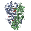
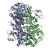
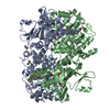
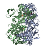
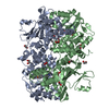
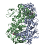
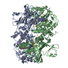
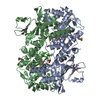
| Title | Crystal Structure of human NAMPT in complex with NVP-LVR596 | ||||||
 Components Components | Nicotinamide phosphoribosyltransferase | ||||||
 Keywords Keywords | TRANSFERASE | ||||||
| Function / homology |  Function and homology information Function and homology informationnicotinamide phosphoribosyltransferase / nicotinamide phosphoribosyltransferase activity / nicotinamide metabolic process / NAD+ biosynthetic process via the salvage pathway / nicotinate metabolic process / NADP+ biosynthetic process / NAD+ biosynthetic process / Nicotinate metabolism / NPAS4 regulates expression of target genes / BMAL1:CLOCK,NPAS2 activates circadian expression ...nicotinamide phosphoribosyltransferase / nicotinamide phosphoribosyltransferase activity / nicotinamide metabolic process / NAD+ biosynthetic process via the salvage pathway / nicotinate metabolic process / NADP+ biosynthetic process / NAD+ biosynthetic process / Nicotinate metabolism / NPAS4 regulates expression of target genes / BMAL1:CLOCK,NPAS2 activates circadian expression / cytokine activity / circadian regulation of gene expression / cell junction / cell-cell signaling / positive regulation of ERK1 and ERK2 cascade / positive regulation of canonical NF-kappaB signal transduction / nuclear speck / mitochondrial matrix / inflammatory response / positive regulation of cell population proliferation / positive regulation of gene expression / signal transduction / positive regulation of transcription by RNA polymerase II / extracellular exosome / identical protein binding / cytosol Similarity search - Function | ||||||
| Biological species |  Homo sapiens (human) Homo sapiens (human) | ||||||
| Method |  X-RAY DIFFRACTION / SYNCHROTRON / FOURIER SYNTHESIS / Resolution: 2.44 Å X-RAY DIFFRACTION / SYNCHROTRON / FOURIER SYNTHESIS / Resolution: 2.44 Å | ||||||
 Authors Authors | Weihofen, W.A. / Thigale, S. | ||||||
 Citation Citation | Journal: To Be Published Title: Identification and structure based design of cellularly active cyclo-propyl carboxamide Nicotinamide phosphoribosyltransferase (NAMPT) inhibitors Authors: Palacios, D. / Meredith, E. / Kawanami, T. / Adams, C. / Chen, X. / Darsigny, V. / Geno, E. / Palermo, M. / Guy, C. / Hewett, J. / Tierney, L. / THigale, S. / Weihofen, W.A. / Agrikar, U. / ...Authors: Palacios, D. / Meredith, E. / Kawanami, T. / Adams, C. / Chen, X. / Darsigny, V. / Geno, E. / Palermo, M. / Guy, C. / Hewett, J. / Tierney, L. / THigale, S. / Weihofen, W.A. / Agrikar, U. / Boynton, G. / George, E. / Wang, L. | ||||||
| History |
|
- Structure visualization
| Structure viewer | Molecule: MolmilJmol/JSmol |
|---|
- Downloads & links
-Download
| PDBx/mmCIF format | 6b76.cif.gz | 399.1 KB | Display | PDBx/mmCIF format |
|---|---|---|---|---|
| PDB format | pdb6b76.ent.gz | 324.5 KB | Display | PDB format |
| PDBx/mmJSON format | 6b76.json.gz | Tree view | PDBx/mmJSON format | |
| Others |  Other downloads Other downloads |
-Validation report
| Arichive directory | https://data.pdbj.org/pub/pdb/validation_reports/b7/6b76ftp://data.pdbj.org/pub/pdb/validation_reports/b7/6b76 | HTTPS FTP |
|---|
-Related structure data
| Related structure data |  6atbSC  6azjC  6b75C S: Starting model for refinement C: citing same article ( |
|---|---|
| Similar structure data |


-Links
 PDBj
PDBj- Assembly
| Deposited unit | 
| ||||||||
|---|---|---|---|---|---|---|---|---|---|
| 1 | 
| ||||||||
| 2 | 
| ||||||||
| Unit cell |
|
-Components
| #1: Protein | Mass: 56942.375 Da / Num. of mol.: 4 Source method: isolated from a genetically manipulated source Source: (gene. exp.) Homo sapiens (human) / Gene: NAMPT, PBEF, PBEF1 / Plasmid: pET41 / Production host:  References: UniProt: P43490, nicotinamide phosphoribosyltransferase #2: Chemical | ChemComp-CVJ / (   Mass: 337.416 Da / Num. of mol.: 4 / Source method: obtained synthetically / Formula: C20H23N3O2 / Feature type: SUBJECT OF INVESTIGATION Mass: 337.416 Da / Num. of mol.: 4 / Source method: obtained synthetically / Formula: C20H23N3O2 / Feature type: SUBJECT OF INVESTIGATION#3: Chemical | ChemComp-PO4 /   Mass: 94.971 Da / Num. of mol.: 11 / Source method: obtained synthetically / Formula: PO4 Mass: 94.971 Da / Num. of mol.: 11 / Source method: obtained synthetically / Formula: PO4#4: Water | ChemComp-HOH / |  Mass: 18.015 Da / Num. of mol.: 874 / Source method: isolated from a natural source / Formula: H2O Mass: 18.015 Da / Num. of mol.: 874 / Source method: isolated from a natural source / Formula: H2O |
|---|
-Experimental details
-Experiment
| Experiment | Method: X-RAY DIFFRACTION / Number of used crystals: 1 |
|---|
- Sample preparation
| Crystal | Density Matthews: 2.37 Å3/Da / Density % sol: 48.14 % |
|---|---|
| Crystal grow | Temperature: 293 K / Method: vapor diffusion, sitting drop / pH: 8 Details: 25 % (w/v) PEG 3350, 200 mM NaCl, 100 mM Tris/HCl pH 8.0 |
-Data collection
| Diffraction | Mean temperature: 100 K |
|---|---|
| Diffraction source | Source: SYNCHROTRON / Site: APS  / Beamline: 17-ID / Wavelength: 1 Å / Beamline: 17-ID / Wavelength: 1 Å |
| Detector | Type: DECTRIS PILATUS3 6M / Detector: PIXEL / Date: Oct 3, 2013 |
| Radiation | Protocol: SINGLE WAVELENGTH / Monochromatic (M) / Laue (L): M / Scattering type: x-ray |
| Radiation wavelength | Wavelength: 1 Å / Relative weight: 1 |
| Reflection | Resolution: 2.44→248.3 Å / Num. obs: 81287 / % possible obs: 100 % / Redundancy: 6.6 % / Biso Wilson estimate: 61.26 Å2 / Rmerge(I) obs: 0.065 / Net I/σ(I): 18.4 |
| Reflection shell | Resolution: 2.44→2.57 Å / Redundancy: 6.6 % / Rmerge(I) obs: 0.582 / Mean I/σ(I) obs: 2.6 / Num. unique all: 11185 / % possible all: 100 |
- Processing
| Software |
| ||||||||||||||||||||||||||||||||||||||||||||||||||||||||||||||||||||||||||||||||||||||||||||||||||||||||||||||||||
|---|---|---|---|---|---|---|---|---|---|---|---|---|---|---|---|---|---|---|---|---|---|---|---|---|---|---|---|---|---|---|---|---|---|---|---|---|---|---|---|---|---|---|---|---|---|---|---|---|---|---|---|---|---|---|---|---|---|---|---|---|---|---|---|---|---|---|---|---|---|---|---|---|---|---|---|---|---|---|---|---|---|---|---|---|---|---|---|---|---|---|---|---|---|---|---|---|---|---|---|---|---|---|---|---|---|---|---|---|---|---|---|---|---|---|---|
| Refinement | Method to determine structure: FOURIER SYNTHESIS Starting model: 6atb Resolution: 2.44→124.15 Å / Cor.coef. Fo:Fc: 0.9363 / Cor.coef. Fo:Fc free: 0.9175 / SU R Cruickshank DPI: 0.424 / Cross valid method: THROUGHOUT / σ(F): 0 / SU R Blow DPI: 0.457 / SU Rfree Blow DPI: 0.252 / SU Rfree Cruickshank DPI: 0.252
| ||||||||||||||||||||||||||||||||||||||||||||||||||||||||||||||||||||||||||||||||||||||||||||||||||||||||||||||||||
| Displacement parameters | Biso mean: 58.39 Å2
| ||||||||||||||||||||||||||||||||||||||||||||||||||||||||||||||||||||||||||||||||||||||||||||||||||||||||||||||||||
| Refine analyze | Luzzati coordinate error obs: 0.284 Å | ||||||||||||||||||||||||||||||||||||||||||||||||||||||||||||||||||||||||||||||||||||||||||||||||||||||||||||||||||
| Refinement step | Cycle: LAST / Resolution: 2.44→124.15 Å
| ||||||||||||||||||||||||||||||||||||||||||||||||||||||||||||||||||||||||||||||||||||||||||||||||||||||||||||||||||
| Refine LS restraints |
| ||||||||||||||||||||||||||||||||||||||||||||||||||||||||||||||||||||||||||||||||||||||||||||||||||||||||||||||||||
| LS refinement shell | Resolution: 2.44→2.5 Å / Total num. of bins used: 20
|
