 Movie
Movie Controller

[English] 日本語
 Yorodumi
- PDB-2bcj: Crystal Structure of G Protein-Coupled Receptor Kinase 2 in Compl... -
+ Open data

- Basic information
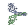
| Entry | Database: PDB / ID: 2bcj | ||||||
|---|---|---|---|---|---|---|---|
| Title | Crystal Structure of G Protein-Coupled Receptor Kinase 2 in Complex with Galpha-q and Gbetagamma Subunits | ||||||
 Components Components |
| ||||||
 Keywords Keywords | TRANSFERASE/HYDROLASE / Peripheral membrane complex / protein kinase / RGS domain / WD40 protein / heterotrimeric G protein / TRANSFERASE-HYDROLASE COMPLEX | ||||||
| Function / homology |  Function and homology information Function and homology informationFatty Acids bound to GPR40 (FFAR1) regulate insulin secretion / PLC beta mediated events / Acetylcholine regulates insulin secretion / forebrain neuron development / Calmodulin induced events / regulation of melanocyte differentiation / negative regulation of the force of heart contraction by chemical signal / negative regulation of relaxation of smooth muscle / beta-adrenergic-receptor kinase / Cooperation of PDCL (PhLP1) and TRiC/CCT in G-protein beta folding ...Fatty Acids bound to GPR40 (FFAR1) regulate insulin secretion / PLC beta mediated events / Acetylcholine regulates insulin secretion / forebrain neuron development / Calmodulin induced events / regulation of melanocyte differentiation / negative regulation of the force of heart contraction by chemical signal / negative regulation of relaxation of smooth muscle / beta-adrenergic-receptor kinase / Cooperation of PDCL (PhLP1) and TRiC/CCT in G-protein beta folding / Edg-2 lysophosphatidic acid receptor binding / Activation of SMO / Thromboxane signalling through TP receptor / adenylate cyclase regulator activity / Extra-nuclear estrogen signaling / alpha-2A adrenergic receptor binding / beta-adrenergic receptor kinase activity / Thrombin signalling through proteinase activated receptors (PARs) / adenylate cyclase-activating G protein-coupled cAMP receptor signaling pathway / G protein-coupled receptor kinase activity / Adenylate cyclase inhibitory pathway / Turbulent (oscillatory, disturbed) flow shear stress activates signaling by PIEZO1 and integrins in endothelial cells / G-protein activation / tachykinin receptor signaling pathway / G alpha (q) signalling events / phospholipase C-activating G protein-coupled acetylcholine receptor signaling pathway / negative regulation of striated muscle contraction / endothelin receptor signaling pathway / Olfactory Signaling Pathway / Sensory perception of sweet, bitter, and umami (glutamate) taste / Synthesis, secretion, and inactivation of Glucagon-like Peptide-1 (GLP-1) / developmental pigmentation / phospholipase C-activating G protein-coupled glutamate receptor signaling pathway / phospholipase C-activating dopamine receptor signaling pathway / Cargo recognition for clathrin-mediated endocytosis / positive regulation of protein localization to cilium / ADP signalling through P2Y purinoceptor 1 / High laminar flow shear stress activates signaling by PIEZO1 and PECAM1:CDH5:KDR in endothelial cells / phospholipase C-activating serotonin receptor signaling pathway / regulation of platelet activation / cytoplasmic side of mitochondrial outer membrane / cranial skeletal system development / Activation of the phototransduction cascade / regulation of the force of heart contraction / desensitization of G protein-coupled receptor signaling pathway / positive regulation of smoothened signaling pathway / maternal behavior / negative regulation of synaptic transmission / G protein-coupled receptor internalization / Adrenaline,noradrenaline inhibits insulin secretion / ADP signalling through P2Y purinoceptor 12 / GTPase activating protein binding / regulation of canonical Wnt signaling pathway / G alpha (i) signalling events / Activation of G protein gated Potassium channels / G-protein activation / G beta:gamma signalling through PI3Kgamma / Prostacyclin signalling through prostacyclin receptor / G beta:gamma signalling through PLC beta / ADP signalling through P2Y purinoceptor 1 / Thromboxane signalling through TP receptor / Presynaptic function of Kainate receptors / G beta:gamma signalling through CDC42 / Inhibition of voltage gated Ca2+ channels via Gbeta/gamma subunits / G alpha (12/13) signalling events / Glucagon-type ligand receptors / G beta:gamma signalling through BTK / ADP signalling through P2Y purinoceptor 12 / Adrenaline,noradrenaline inhibits insulin secretion / embryonic digit morphogenesis / Cooperation of PDCL (PhLP1) and TRiC/CCT in G-protein beta folding / Ca2+ pathway / G alpha (z) signalling events / Thrombin signalling through proteinase activated receptors (PARs) / Extra-nuclear estrogen signaling / G alpha (s) signalling events / G alpha (q) signalling events / glutamate receptor signaling pathway / G alpha (i) signalling events / Glucagon-like Peptide-1 (GLP1) regulates insulin secretion / neuron remodeling / ligand-gated ion channel signaling pathway / High laminar flow shear stress activates signaling by PIEZO1 and PECAM1:CDH5:KDR in endothelial cells / neurotransmitter receptor localization to postsynaptic specialization membrane / Vasopressin regulates renal water homeostasis via Aquaporins / alkylglycerophosphoethanolamine phosphodiesterase activity / negative regulation of potassium ion transport / action potential / regulation of signal transduction / postsynaptic cytosol / adenylate cyclase-activating adrenergic receptor signaling pathway / cellular response to acidic pH / enzyme regulator activity / cardiac muscle contraction / adenylate cyclase inhibitor activity / hormone-mediated signaling pathway / positive regulation of protein localization to cell cortex / T cell migration / D2 dopamine receptor binding / adenylate cyclase-inhibiting serotonin receptor signaling pathway Similarity search - Function | ||||||
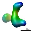
| Biological species |  | ||||||
| Method |  X-RAY DIFFRACTION / SYNCHROTRON / MOLECULAR REPLACEMENT / Resolution: 3.061 Å X-RAY DIFFRACTION / SYNCHROTRON / MOLECULAR REPLACEMENT / Resolution: 3.061 Å | ||||||
 Authors Authors | Tesmer, J.J.G. | ||||||
 Citation Citation | Journal: Science / Year: 2005 Title: Snapshot of Activated G Proteins at the Membrane: The Gq-GRK2-G Complex Authors: Tesmer, V.M. / Kawano, T. / Shankaranarayanan, A. / Kozasa, T. / Tesmer, J.J.G. | ||||||
| History |
|
- Structure visualization
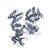
| Structure viewer | Molecule: MolmilJmol/JSmol |
|---|
- Downloads & links
-Download
| PDBx/mmCIF format | 2bcj.cif.gz | 283.6 KB | Display | PDBx/mmCIF format |
|---|---|---|---|---|
| PDB format | pdb2bcj.ent.gz | 221.8 KB | Display | PDB format |
| PDBx/mmJSON format | 2bcj.json.gz | Tree view | PDBx/mmJSON format | |
| Others |  Other downloads Other downloads |
-Validation report
| Arichive directory | https://data.pdbj.org/pub/pdb/validation_reports/bc/2bcjftp://data.pdbj.org/pub/pdb/validation_reports/bc/2bcj | HTTPS FTP |
|---|
-Related structure data
| Related structure data | 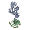 1omwS S: Starting model for refinement |
|---|---|
| Similar structure data |


-Links
 PDBj
PDBj


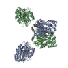
- Assembly
| Deposited unit | 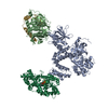
| ||||||||
|---|---|---|---|---|---|---|---|---|---|
| 1 |
| ||||||||
| Unit cell |
|
-Components
-Protein , 1 types, 1 molecules A
| #1: Protein | Mass: 79749.922 Da / Num. of mol.: 1 / Fragment: residues 28-689 / Mutation: S670A Source method: isolated from a genetically manipulated source Source: (gene. exp.)  Trichoplusia ni (cabbage looper) Trichoplusia ni (cabbage looper)References: UniProt: P21146, beta-adrenergic-receptor kinase |
|---|
-Guanine nucleotide-binding protein ... , 3 types, 3 molecules BGQ
| #2: Protein | Mass: 37311.770 Da / Num. of mol.: 1 Source method: isolated from a genetically manipulated source Source: (gene. exp.) Trichoplusia ni (cabbage looper) / References: UniProt: P62871 |
|---|---|
| #3: Protein | Mass: 7845.078 Da / Num. of mol.: 1 / Mutation: C68S Source method: isolated from a genetically manipulated source Source: (gene. exp.) Trichoplusia ni (cabbage looper) / References: UniProt: P63212 |
| #4: Protein | Mass: 41298.895 Da / Num. of mol.: 1 Source method: isolated from a genetically manipulated source Source: (gene. exp.) Genus: Rattus, Mus / Species: , / Gene: Gnai1, Gnai-1, Gnaq / Plasmid: pFastBacHTA / Cell line (production host): High-5 cells / Production host: Trichoplusia ni (cabbage looper) / References: UniProt: P10824, UniProt: P21279 |
-Non-polymers , 4 types, 8 molecules 


| #5: Chemical | ChemComp-MG /  Mass: 24.305 Da / Num. of mol.: 1 / Source method: obtained synthetically / Formula: Mg Mass: 24.305 Da / Num. of mol.: 1 / Source method: obtained synthetically / Formula: Mg |
|---|---|
| #6: Chemical | ChemComp-ALF /  Mass: 102.975 Da / Num. of mol.: 1 / Source method: obtained synthetically / Formula: AlF4 Mass: 102.975 Da / Num. of mol.: 1 / Source method: obtained synthetically / Formula: AlF4 |
| #7: Chemical | ChemComp-GDP /  Type: RNA linking / Mass: 443.201 Da / Num. of mol.: 1 / Source method: obtained synthetically / Formula: C10H15N5O11P2 / Comment: GDP, energy-carrying molecule*YM Type: RNA linking / Mass: 443.201 Da / Num. of mol.: 1 / Source method: obtained synthetically / Formula: C10H15N5O11P2 / Comment: GDP, energy-carrying molecule*YM |
| #8: Water | ChemComp-HOH / Mass: 18.015 Da / Num. of mol.: 5 / Source method: isolated from a natural source / Formula: H2O |
-Details
| Has protein modification | Y |
|---|
-Experimental details
-Experiment
| Experiment | Method: X-RAY DIFFRACTION / Number of used crystals: 1 |
|---|
- Sample preparation
| Crystal | Density Matthews: 3.22 Å3/Da / Density % sol: 61.81 % |
|---|---|
| Crystal grow | Temperature: 277 K / Method: vapor diffusion, hanging drop / pH: 6.5 Details: 9% PEG8000, 1M NaCl, 0.1M MES, pH 6.5, VAPOR DIFFUSION, HANGING DROP, temperature 277K |
-Data collection
| Diffraction | Mean temperature: 100 K |
|---|---|
| Diffraction source | Source: SYNCHROTRON / Site: ALS  / Beamline: 8.3.1 / Wavelength: 1.116 Å / Beamline: 8.3.1 / Wavelength: 1.116 Å |
| Detector | Type: ADSC QUANTUM 4 / Detector: CCD / Date: Apr 7, 2005 |
| Radiation | Monochromator: Si(111) / Protocol: SINGLE WAVELENGTH / Monochromatic (M) / Laue (L): M / Scattering type: x-ray |
| Radiation wavelength | Wavelength: 1.116 Å / Relative weight: 1 |
| Reflection | Resolution: 3.06→46.78 Å / Num. all: 37836 / Num. obs: 37836 / % possible obs: 100 % / Observed criterion σ(F): 0 / Observed criterion σ(I): -2 / Redundancy: 5.7 % / Biso Wilson estimate: 75 Å2 / Rsym value: 0.133 / Net I/σ(I): 13.55 |
| Reflection shell | Resolution: 3.06→3.21 Å / Mean I/σ(I) obs: 1.1 / Num. unique all: 3738 / Rsym value: 0.849 / % possible all: 99.9 |
- Processing
| Software |
| ||||||||||||||||||||||||||||||||||||||||||||||||||||||||||||||||||||||||||||||||||||||||||||||||||||||||||||||||||||||||||||||||||
|---|---|---|---|---|---|---|---|---|---|---|---|---|---|---|---|---|---|---|---|---|---|---|---|---|---|---|---|---|---|---|---|---|---|---|---|---|---|---|---|---|---|---|---|---|---|---|---|---|---|---|---|---|---|---|---|---|---|---|---|---|---|---|---|---|---|---|---|---|---|---|---|---|---|---|---|---|---|---|---|---|---|---|---|---|---|---|---|---|---|---|---|---|---|---|---|---|---|---|---|---|---|---|---|---|---|---|---|---|---|---|---|---|---|---|---|---|---|---|---|---|---|---|---|---|---|---|---|---|---|---|---|
| Refinement | Method to determine structure: MOLECULAR REPLACEMENT Starting model: PDB ENTRY 1OMW, homology model of Galphaq Resolution: 3.061→46.78 Å / Cor.coef. Fo:Fc: 0.943 / SU B: 50.535 / SU ML: 0.343 / TLS residual ADP flag: LIKELY RESIDUAL / Isotropic thermal model: isotropic / σ(F): 0 / Stereochemistry target values: MAXIMUM LIKELIHOOD Details: HYDROGENS HAVE BEEN ADDED IN THE RIDING POSITIONS. Rfree was not used for the last three rounds of refinement.
| ||||||||||||||||||||||||||||||||||||||||||||||||||||||||||||||||||||||||||||||||||||||||||||||||||||||||||||||||||||||||||||||||||
| Solvent computation | Ion probe radii: 0.8 Å / Shrinkage radii: 0.8 Å / VDW probe radii: 1.2 Å / Solvent model: MASK | ||||||||||||||||||||||||||||||||||||||||||||||||||||||||||||||||||||||||||||||||||||||||||||||||||||||||||||||||||||||||||||||||||
| Displacement parameters | Biso mean: 99.74 Å2
| ||||||||||||||||||||||||||||||||||||||||||||||||||||||||||||||||||||||||||||||||||||||||||||||||||||||||||||||||||||||||||||||||||
| Refinement step | Cycle: LAST / Resolution: 3.061→46.78 Å
| ||||||||||||||||||||||||||||||||||||||||||||||||||||||||||||||||||||||||||||||||||||||||||||||||||||||||||||||||||||||||||||||||||
| Refine LS restraints |
| ||||||||||||||||||||||||||||||||||||||||||||||||||||||||||||||||||||||||||||||||||||||||||||||||||||||||||||||||||||||||||||||||||
| LS refinement shell | Resolution: 3.061→3.141 Å / Total num. of bins used: 20
| ||||||||||||||||||||||||||||||||||||||||||||||||||||||||||||||||||||||||||||||||||||||||||||||||||||||||||||||||||||||||||||||||||
| Refinement TLS params. | Method: refined / Origin x: 16.358 Å / Origin y: -1.5957 Å / Origin z: 6.6176 Å
| ||||||||||||||||||||||||||||||||||||||||||||||||||||||||||||||||||||||||||||||||||||||||||||||||||||||||||||||||||||||||||||||||||
| Refinement TLS group | Refine-ID: X-RAY DIFFRACTION / Refine TLS-ID: 1 / Selection: ALL
|
