 Movie
Movie Controller

[English] 日本語
 Yorodumi
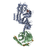
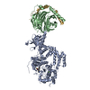
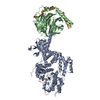
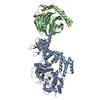
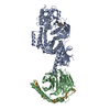
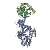
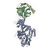
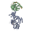
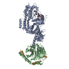
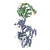
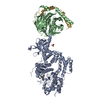
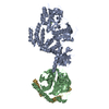
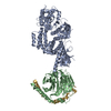
- PDB-1omw: Crystal Structure of the complex between G Protein-Coupled Recept... -
+ Open data

- Basic information
| Entry | Database: PDB / ID: 1omw | ||||||
|---|---|---|---|---|---|---|---|
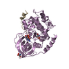
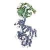
| Title | Crystal Structure of the complex between G Protein-Coupled Receptor Kinase 2 and Heterotrimeric G Protein beta 1 and gamma 2 subunits | ||||||
 Components Components |
| ||||||
 Keywords Keywords | TRANSFERASE / WD-40 repeat | ||||||
| Function / homology |  Function and homology information Function and homology informationCalmodulin induced events / negative regulation of the force of heart contraction by chemical signal / negative regulation of relaxation of smooth muscle / beta-adrenergic-receptor kinase / Edg-2 lysophosphatidic acid receptor binding / Activation of SMO / alpha-2A adrenergic receptor binding / beta-adrenergic receptor kinase activity / G protein-coupled receptor kinase activity / tachykinin receptor signaling pathway ...Calmodulin induced events / negative regulation of the force of heart contraction by chemical signal / negative regulation of relaxation of smooth muscle / beta-adrenergic-receptor kinase / Edg-2 lysophosphatidic acid receptor binding / Activation of SMO / alpha-2A adrenergic receptor binding / beta-adrenergic receptor kinase activity / G protein-coupled receptor kinase activity / tachykinin receptor signaling pathway / negative regulation of striated muscle contraction / Olfactory Signaling Pathway / Sensory perception of sweet, bitter, and umami (glutamate) taste / Synthesis, secretion, and inactivation of Glucagon-like Peptide-1 (GLP-1) / Cargo recognition for clathrin-mediated endocytosis / positive regulation of protein localization to cilium / cytoplasmic side of mitochondrial outer membrane / Activation of the phototransduction cascade / regulation of the force of heart contraction / desensitization of G protein-coupled receptor signaling pathway / positive regulation of smoothened signaling pathway / G protein-coupled receptor internalization / Activation of G protein gated Potassium channels / G-protein activation / G beta:gamma signalling through PI3Kgamma / Prostacyclin signalling through prostacyclin receptor / G beta:gamma signalling through PLC beta / ADP signalling through P2Y purinoceptor 1 / Thromboxane signalling through TP receptor / Presynaptic function of Kainate receptors / G beta:gamma signalling through CDC42 / Inhibition of voltage gated Ca2+ channels via Gbeta/gamma subunits / G alpha (12/13) signalling events / Glucagon-type ligand receptors / G beta:gamma signalling through BTK / ADP signalling through P2Y purinoceptor 12 / Adrenaline,noradrenaline inhibits insulin secretion / Cooperation of PDCL (PhLP1) and TRiC/CCT in G-protein beta folding / Ca2+ pathway / G alpha (z) signalling events / Thrombin signalling through proteinase activated receptors (PARs) / Extra-nuclear estrogen signaling / G alpha (s) signalling events / G alpha (q) signalling events / Glucagon-like Peptide-1 (GLP1) regulates insulin secretion / G alpha (i) signalling events / High laminar flow shear stress activates signaling by PIEZO1 and PECAM1:CDH5:KDR in endothelial cells / Vasopressin regulates renal water homeostasis via Aquaporins / regulation of signal transduction / adenylate cyclase-activating adrenergic receptor signaling pathway / cardiac muscle contraction / viral genome replication / intracellular protein transport / G protein-coupled receptor binding / G protein-coupled acetylcholine receptor signaling pathway / photoreceptor disc membrane / cellular response to catecholamine stimulus / adenylate cyclase-activating dopamine receptor signaling pathway / cellular response to prostaglandin E stimulus / heterotrimeric G-protein complex / G-protein beta-subunit binding / heart development / sensory perception of taste / presynapse / signaling receptor complex adaptor activity / retina development in camera-type eye / GTPase binding / phospholipase C-activating G protein-coupled receptor signaling pathway / protein kinase activity / cell population proliferation / postsynapse / G protein-coupled receptor signaling pathway / GTPase activity / symbiont entry into host cell / synapse / protein-containing complex binding / ATP binding / membrane / plasma membrane / cytoplasm / cytosol Similarity search - Function | ||||||
| Biological species |  | ||||||
| Method |  X-RAY DIFFRACTION / SYNCHROTRON / MOLECULAR REPLACEMENT / Resolution: 2.5 Å X-RAY DIFFRACTION / SYNCHROTRON / MOLECULAR REPLACEMENT / Resolution: 2.5 Å | ||||||
 Authors Authors | Lodowski, D.T. / Pitcher, J.A. / Capel, W.D. / Lefkowitz, R.J. / Tesmer, J.J.G. | ||||||
 Citation Citation | Journal: Science / Year: 2003 Title: Keeping G proteins at Bay: A Complex Between G Protein-Coupled Receptor Kinase 2 and G-Beta-Gamma Authors: Lodowski, D.T. / Pitcher, J.A. / Capel, W.D. / Lefkowitz, R.J. / Tesmer, J.J.G. | ||||||
| History |
|
- Structure visualization
| Structure viewer | Molecule: MolmilJmol/JSmol |
|---|
- Downloads & links
-Download
| PDBx/mmCIF format | 1omw.cif.gz | 213.6 KB | Display | PDBx/mmCIF format |
|---|---|---|---|---|
| PDB format | pdb1omw.ent.gz | 169.6 KB | Display | PDB format |
| PDBx/mmJSON format | 1omw.json.gz | Tree view | PDBx/mmJSON format | |
| Others |  Other downloads Other downloads |
-Validation report
| Arichive directory | https://data.pdbj.org/pub/pdb/validation_reports/om/1omwftp://data.pdbj.org/pub/pdb/validation_reports/om/1omw | HTTPS FTP |
|---|
-Related structure data


-Links
 PDBj
PDBj


- Assembly
| Deposited unit | 
| ||||||||
|---|---|---|---|---|---|---|---|---|---|
| 1 |
| ||||||||
| Unit cell |
|
-Components
| #1: Protein | Mass: 79749.922 Da / Num. of mol.: 1 / Mutation: S670A Source method: isolated from a genetically manipulated source Source: (gene. exp.)   Spodoptera frugiperda (fall armyworm) / Strain (production host): Sf9 / References: UniProt: P21146, EC: 2.7.1.126 Spodoptera frugiperda (fall armyworm) / Strain (production host): Sf9 / References: UniProt: P21146, EC: 2.7.1.126 |
|---|---|
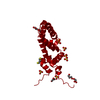
| #2: Protein | Mass: 37416.930 Da / Num. of mol.: 1 Source method: isolated from a genetically manipulated source Source: (gene. exp.) Spodoptera frugiperda (fall armyworm) / Strain (production host): SF9 / References: UniProt: P62871 |
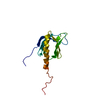
| #3: Protein | Mass: 8406.658 Da / Num. of mol.: 1 Source method: isolated from a genetically manipulated source Source: (gene. exp.) Spodoptera frugiperda (fall armyworm) / Strain (production host): Sf9 / References: UniProt: P63212 |
| #4: Water | ChemComp-HOH /  Mass: 18.015 Da / Num. of mol.: 26 / Source method: isolated from a natural source / Formula: H2O Mass: 18.015 Da / Num. of mol.: 26 / Source method: isolated from a natural source / Formula: H2O |
-Experimental details
-Experiment
| Experiment | Method: X-RAY DIFFRACTION / Number of used crystals: 1 |
|---|
- Sample preparation
| Crystal | Density Matthews: 3 Å3/Da / Density % sol: 59.3 % | |||||||||||||||||||||||||||||||||||||||||||||||||
|---|---|---|---|---|---|---|---|---|---|---|---|---|---|---|---|---|---|---|---|---|---|---|---|---|---|---|---|---|---|---|---|---|---|---|---|---|---|---|---|---|---|---|---|---|---|---|---|---|---|---|
| Crystal grow | Temperature: 277 K / Method: vapor diffusion, hanging drop / pH: 5.75 Details: PEG 3350, Sodium Chloride, CHAPS, ATP, Magnesium Chloride, Mastoparan , MES pH 5.75, VAPOR DIFFUSION, HANGING DROP, temperature 277K | |||||||||||||||||||||||||||||||||||||||||||||||||
| Crystal grow | *PLUS Method: vapor diffusion, hanging dropDetails: Lodowski, D.T., (2003) Acta Crystallogr., D59, 936. | |||||||||||||||||||||||||||||||||||||||||||||||||
| Components of the solutions | *PLUS
|
-Data collection
| Diffraction |
| ||||||||||||||||||
|---|---|---|---|---|---|---|---|---|---|---|---|---|---|---|---|---|---|---|---|
| Diffraction source |
| ||||||||||||||||||
| Detector |
| ||||||||||||||||||
| Radiation |
| ||||||||||||||||||
| Radiation wavelength |
| ||||||||||||||||||
| Reflection | Resolution: 2.5→20 Å / Num. all: 135084 / Num. obs: 56160 / % possible obs: 73.6 % / Observed criterion σ(F): -2 / Observed criterion σ(I): -2 / Redundancy: 2.4 % / Biso Wilson estimate: 37.42 Å2 / Rsym value: 0.05 / Net I/σ(I): 15.3 | ||||||||||||||||||
| Reflection shell | Resolution: 2.5→2.59 Å / Redundancy: 0.67 % / Mean I/σ(I) obs: 2.7 / Num. unique all: 1977 / Rsym value: 0.253 / % possible all: 12.7 | ||||||||||||||||||
| Reflection | *PLUS Num. obs: 38258 / Rmerge(I) obs: 0.053 | ||||||||||||||||||
| Reflection shell | *PLUS % possible obs: 12.7 % / Rmerge(I) obs: 0.253 |

- Processing
| Software |
| ||||||||||||||||||||||||||||||||||||||||||||||||||||||||||||||||||||||||||||||||||||||||||||||||||||||||||||||||||||||||||||||||||||||||||||||||||||||
|---|---|---|---|---|---|---|---|---|---|---|---|---|---|---|---|---|---|---|---|---|---|---|---|---|---|---|---|---|---|---|---|---|---|---|---|---|---|---|---|---|---|---|---|---|---|---|---|---|---|---|---|---|---|---|---|---|---|---|---|---|---|---|---|---|---|---|---|---|---|---|---|---|---|---|---|---|---|---|---|---|---|---|---|---|---|---|---|---|---|---|---|---|---|---|---|---|---|---|---|---|---|---|---|---|---|---|---|---|---|---|---|---|---|---|---|---|---|---|---|---|---|---|---|---|---|---|---|---|---|---|---|---|---|---|---|---|---|---|---|---|---|---|---|---|---|---|---|---|---|---|---|
| Refinement | Method to determine structure: MOLECULAR REPLACEMENT Starting model: PDB ENTRY 1ABG, 1ATP, 1BAK, 1DK8 Resolution: 2.5→20 Å / Cor.coef. Fo:Fc: 0.95 / SU B: 10.266 / SU ML: 0.205 / TLS residual ADP flag: LIKELY RESIDUAL / Isotropic thermal model: TLS refinement Cross valid method: The structure was initally solved with R-free as the cross validation method, but for the last three rounds of refinement, the structure was refined against all of the data. σ(F): -2 / ESU R: 0.72 / Stereochemistry target values: MAXIMUM LIKELIHOOD Details: HYDROGENS HAVE BEEN ADDED IN THE RIDING POSITIONS. Due to the extreme anisotropy of the data, a three dimensional ellipsoid with resolution limits corresponding to the maximum diffraction in ...Details: HYDROGENS HAVE BEEN ADDED IN THE RIDING POSITIONS. Due to the extreme anisotropy of the data, a three dimensional ellipsoid with resolution limits corresponding to the maximum diffraction in each direction was defined to select reflections used in refinement. These diffraction limits were 2.4, 2.8, 3.0 Angstroms. The 2.4 A direction corresponds to 37.9 degrees inclined from the a* axis, the 2.8 A direction corresponds to the b* axis and the 3.0 A direction corresponds to direction 1 X b*.
| ||||||||||||||||||||||||||||||||||||||||||||||||||||||||||||||||||||||||||||||||||||||||||||||||||||||||||||||||||||||||||||||||||||||||||||||||||||||
| Solvent computation | Ion probe radii: 0.8 Å / Shrinkage radii: 0.8 Å / VDW probe radii: 1.4 Å / Solvent model: BABINET MODEL WITH MASK | ||||||||||||||||||||||||||||||||||||||||||||||||||||||||||||||||||||||||||||||||||||||||||||||||||||||||||||||||||||||||||||||||||||||||||||||||||||||
| Displacement parameters | Biso mean: 27.933 Å2
| ||||||||||||||||||||||||||||||||||||||||||||||||||||||||||||||||||||||||||||||||||||||||||||||||||||||||||||||||||||||||||||||||||||||||||||||||||||||
| Refinement step | Cycle: LAST / Resolution: 2.5→20 Å
| ||||||||||||||||||||||||||||||||||||||||||||||||||||||||||||||||||||||||||||||||||||||||||||||||||||||||||||||||||||||||||||||||||||||||||||||||||||||
| Refine LS restraints |
| ||||||||||||||||||||||||||||||||||||||||||||||||||||||||||||||||||||||||||||||||||||||||||||||||||||||||||||||||||||||||||||||||||||||||||||||||||||||
| LS refinement shell | Resolution: 2.5→2.564 Å / Total num. of bins used: 20
| ||||||||||||||||||||||||||||||||||||||||||||||||||||||||||||||||||||||||||||||||||||||||||||||||||||||||||||||||||||||||||||||||||||||||||||||||||||||
| Refinement TLS params. | Method: refined / Refine-ID: X-RAY DIFFRACTION
| ||||||||||||||||||||||||||||||||||||||||||||||||||||||||||||||||||||||||||||||||||||||||||||||||||||||||||||||||||||||||||||||||||||||||||||||||||||||
| Refinement TLS group |
| ||||||||||||||||||||||||||||||||||||||||||||||||||||||||||||||||||||||||||||||||||||||||||||||||||||||||||||||||||||||||||||||||||||||||||||||||||||||
| Refinement | *PLUS Highest resolution: 2.5 Å / Lowest resolution: 20 Å / % reflection Rfree: 5.1 % | ||||||||||||||||||||||||||||||||||||||||||||||||||||||||||||||||||||||||||||||||||||||||||||||||||||||||||||||||||||||||||||||||||||||||||||||||||||||
| Solvent computation | *PLUS | ||||||||||||||||||||||||||||||||||||||||||||||||||||||||||||||||||||||||||||||||||||||||||||||||||||||||||||||||||||||||||||||||||||||||||||||||||||||
| Displacement parameters | *PLUS | ||||||||||||||||||||||||||||||||||||||||||||||||||||||||||||||||||||||||||||||||||||||||||||||||||||||||||||||||||||||||||||||||||||||||||||||||||||||
| Refine LS restraints | *PLUS
|
