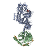
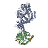
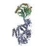
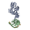
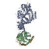
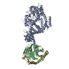
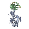
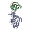
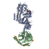
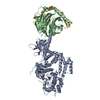
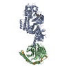
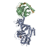
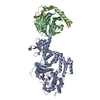
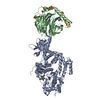
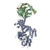
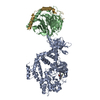
Entry Database : PDB / ID : 3cikTitle Human GRK2 in Complex with Gbetagamma subunits Beta-adrenergic receptor kinase 1 Guanine nucleotide-binding protein G(I)/G(S)/G(O) subunit gamma-2 Guanine nucleotide-binding protein G(I)/G(S)/G(T) subunit beta-1 Keywords / / / / / / / / / / / / / / / / Function / homology Function Domain/homology Component
/ / / / / / / / / / / / / / / / / / / / / / / / / / / / / / / / / / / / / / / / / / / / / / / / / / / / / / / / / / / / / / / / / / / / / / / / / / / / / / / / / / / / / / / / / / / / / / / / / / / / / / / / / / / / / / / / / / / / / / / / / / / / / / / / / / / / / / / / / / / / / / / / / / / / / / / / / / / / / / / / / / / / / Biological species Homo sapiens (human)Bos taurus (domestic cattle)Method / / Resolution : 2.75 Å Authors Tesmer, J.J.G. / Lodowski, D.T. Journal : J.Med.Chem. / Year : 2010Title : Structure of human G protein-coupled receptor kinase 2 in complex with the kinase inhibitor balanol.Authors : Tesmer, J.J. / Tesmer, V.M. / Lodowski, D.T. / Steinhagen, H. / Huber, J. History Deposition Mar 11, 2008 Deposition site / Processing site Revision 1.0 Feb 17, 2009 Provider / Type Revision 1.1 Jul 13, 2011 Group / Version format complianceRevision 1.2 Mar 7, 2012 Group Revision 1.3 Mar 26, 2025 Group Data collection / Database references ... Data collection / Database references / Derived calculations / Structure summary Category chem_comp_atom / chem_comp_bond ... chem_comp_atom / chem_comp_bond / database_2 / pdbx_entry_details / pdbx_modification_feature / struct_conn / struct_ref_seq_dif / struct_site Item _database_2.pdbx_DOI / _database_2.pdbx_database_accession ... _database_2.pdbx_DOI / _database_2.pdbx_database_accession / _struct_conn.pdbx_leaving_atom_flag / _struct_ref_seq_dif.details / _struct_site.pdbx_auth_asym_id / _struct_site.pdbx_auth_comp_id / _struct_site.pdbx_auth_seq_id
Show all Show less
 Movie
Movie Controller


 Open data
 Basic information
 Components
 Keywords
 Function and homology information
 Homo sapiens (human)
 X-RAY DIFFRACTION /
  Authors
 Citation
 Structure visualization
 Downloads & links
 Other downloads


 PDBj
PDBj


 Assembly
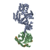

 Spodoptera frugiperda (fall armyworm)
 Mass: 24.305 Da / Num. of mol.: 1 / Source method: obtained synthetically / Formula: Mg
 Mass: 18.015 Da / Num. of mol.: 19 / Source method: isolated from a natural source / Formula: H2O
 Sample preparation
 / Beamline: 19-BM / Wavelength: 1.033 Å
 Processing