 Movie
Movie Controller

[English] 日本語
 Yorodumi
- PDB-7k7z: Structure of a hit for G Protein Coupled Receptor Kinase 2 (GRK2)... -
+ Open data

- Basic information
| Entry | Database: PDB / ID: 7k7z | ||||||
|---|---|---|---|---|---|---|---|
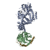
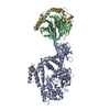
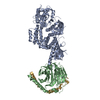
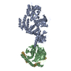
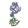
| Title | Structure of a hit for G Protein Coupled Receptor Kinase 2 (GRK2) Inhibitor for the Potential Treatment of Heart Failure | ||||||
 Components Components |
| ||||||
 Keywords Keywords | SIGNALING PROTEIN / TRANSFERASE/INHIBITOR / serine/threonine protein kinase / TRANSFERASE-INHIBITOR complex | ||||||
| Function / homology |  Function and homology information Function and homology informationnegative regulation of the force of heart contraction by chemical signal / negative regulation of relaxation of smooth muscle / beta-adrenergic-receptor kinase / Edg-2 lysophosphatidic acid receptor binding / alpha-2A adrenergic receptor binding / beta-adrenergic receptor kinase activity / G-protein-coupled receptor kinase / G protein-coupled receptor kinase activity / tachykinin receptor signaling pathway / positive regulation of catecholamine secretion ...negative regulation of the force of heart contraction by chemical signal / negative regulation of relaxation of smooth muscle / beta-adrenergic-receptor kinase / Edg-2 lysophosphatidic acid receptor binding / alpha-2A adrenergic receptor binding / beta-adrenergic receptor kinase activity / G-protein-coupled receptor kinase / G protein-coupled receptor kinase activity / tachykinin receptor signaling pathway / positive regulation of catecholamine secretion / negative regulation of striated muscle contraction / Activation of SMO / cytoplasmic side of mitochondrial outer membrane / regulation of the force of heart contraction / desensitization of G protein-coupled receptor signaling pathway / Calmodulin induced events / cardiac muscle contraction / viral genome replication / receptor internalization / G protein-coupled receptor binding / Olfactory Signaling Pathway / Activation of the phototransduction cascade / G protein-coupled acetylcholine receptor signaling pathway / G beta:gamma signalling through PLC beta / Presynaptic function of Kainate receptors / Thromboxane signalling through TP receptor / Activation of G protein gated Potassium channels / Inhibition of voltage gated Ca2+ channels via Gbeta/gamma subunits / G-protein activation / Glucagon signaling in metabolic regulation / G beta:gamma signalling through CDC42 / Prostacyclin signalling through prostacyclin receptor / Synthesis, secretion, and inactivation of Glucagon-like Peptide-1 (GLP-1) / G beta:gamma signalling through BTK / photoreceptor disc membrane / ADP signalling through P2Y purinoceptor 12 / Glucagon-type ligand receptors / Sensory perception of sweet, bitter, and umami (glutamate) taste / Adrenaline,noradrenaline inhibits insulin secretion / Vasopressin regulates renal water homeostasis via Aquaporins / Glucagon-like Peptide-1 (GLP1) regulates insulin secretion / G alpha (z) signalling events / cellular response to catecholamine stimulus / ADP signalling through P2Y purinoceptor 1 / G beta:gamma signalling through PI3Kgamma / ADORA2B mediated anti-inflammatory cytokines production / Cargo recognition for clathrin-mediated endocytosis / adenylate cyclase-activating dopamine receptor signaling pathway / Cooperation of PDCL (PhLP1) and TRiC/CCT in G-protein beta folding / GPER1 signaling / cellular response to prostaglandin E stimulus / heterotrimeric G-protein complex / Inactivation, recovery and regulation of the phototransduction cascade / G alpha (12/13) signalling events / G-protein beta-subunit binding / extracellular vesicle / sensory perception of taste / presynapse / Thrombin signalling through proteinase activated receptors (PARs) / signaling receptor complex adaptor activity / retina development in camera-type eye / fibroblast proliferation / GTPase binding / Ca2+ pathway / High laminar flow shear stress activates signaling by PIEZO1 and PECAM1:CDH5:KDR in endothelial cells / G alpha (i) signalling events / G alpha (s) signalling events / G alpha (q) signalling events / phospholipase C-activating G protein-coupled receptor signaling pathway / Ras protein signal transduction / protein kinase activity / cell population proliferation / cilium / postsynapse / Extra-nuclear estrogen signaling / G protein-coupled receptor signaling pathway / lysosomal membrane / GTPase activity / symbiont entry into host cell / synapse / protein-containing complex binding / signal transduction / extracellular exosome / ATP binding / membrane / plasma membrane / cytosol / cytoplasm Similarity search - Function | ||||||
| Biological species |  Homo sapiens (human) Homo sapiens (human) | ||||||
| Method |  X-RAY DIFFRACTION / SYNCHROTRON / MOLECULAR REPLACEMENT / Resolution: 2.60608692764 Å X-RAY DIFFRACTION / SYNCHROTRON / MOLECULAR REPLACEMENT / Resolution: 2.60608692764 Å | ||||||
 Authors Authors | Spurlino, J.C. / Milligan, C. | ||||||
 Citation Citation | Journal: Bioorg.Med.Chem.Lett. / Year: 2020 Title: Hit-to-lead optimization and discovery of a potent, and orally bioavailable G protein coupled receptor kinase 2 (GRK2) inhibitor. Authors: Xu, G. / Gaul, M.D. / Liu, Z. / DesJarlais, R.L. / Qi, J. / Wang, W. / Krosky, D. / Petrounia, I. / Milligan, C.M. / Hermans, A. / Lu, H.R. / Huang, D.Z. / Xu, J.Z. / Spurlino, J.C. | ||||||
| History |
|
- Structure visualization
| Structure viewer | Molecule: MolmilJmol/JSmol |
|---|
- Downloads & links
-Download
| PDBx/mmCIF format | 7k7z.cif.gz | 505.3 KB | Display | PDBx/mmCIF format |
|---|---|---|---|---|
| PDB format | pdb7k7z.ent.gz | 346.6 KB | Display | PDB format |
| PDBx/mmJSON format | 7k7z.json.gz | Tree view | PDBx/mmJSON format | |
| Others |  Other downloads Other downloads |
-Validation report
| Arichive directory | https://data.pdbj.org/pub/pdb/validation_reports/k7/7k7zftp://data.pdbj.org/pub/pdb/validation_reports/k7/7k7z | HTTPS FTP |
|---|
-Related structure data

-Links
 PDBj
PDBj


- Assembly
| Deposited unit | 
| ||||||||||
|---|---|---|---|---|---|---|---|---|---|---|---|
| 1 |
| ||||||||||
| Unit cell |
|
-Components
| #1: Protein | Mass: 74545.641 Da / Num. of mol.: 1 Source method: isolated from a genetically manipulated source Source: (gene. exp.) Homo sapiens (human) / Gene: GRK2, ADRBK1, BARK, BARK1 / Production host:  unidentified baculovirus unidentified baculovirusReferences: UniProt: P25098, beta-adrenergic-receptor kinase |
|---|---|
| #2: Protein | Mass: 37285.734 Da / Num. of mol.: 1 Source method: isolated from a genetically manipulated source Source: (gene. exp.) Homo sapiens (human) / Gene: GNB1 / Production host: unidentified baculovirus / References: UniProt: P62873 |
| #3: Protein | Mass: 6519.523 Da / Num. of mol.: 1 Source method: isolated from a genetically manipulated source Source: (gene. exp.) Homo sapiens (human) / Gene: GNG2 / Production host: unidentified baculovirus / References: UniProt: P59768 |
| #4: Chemical | ChemComp-W4G /   Mass: 302.330 Da / Num. of mol.: 1 / Source method: obtained synthetically / Formula: C18H14N4O / Feature type: SUBJECT OF INVESTIGATION Mass: 302.330 Da / Num. of mol.: 1 / Source method: obtained synthetically / Formula: C18H14N4O / Feature type: SUBJECT OF INVESTIGATION |
| Has ligand of interest | Y |
-Experimental details
-Experiment
| Experiment | Method: X-RAY DIFFRACTION / Number of used crystals: 1 |
|---|
- Sample preparation
| Crystal | Density Matthews: 3.04 Å3/Da / Density % sol: 59.5 % |
|---|---|
| Crystal grow | Temperature: 293 K / Method: vapor diffusion / pH: 6.5 / Details: 2% PEG 3350, 0.1M MES pH 6.5, 0.2M NaCl |
-Data collection
| Diffraction | Mean temperature: 80 K / Serial crystal experiment: N |
|---|---|
| Diffraction source | Source: SYNCHROTRON / Site: APS  / Beamline: 17-ID / Wavelength: 1 Å / Beamline: 17-ID / Wavelength: 1 Å |
| Detector | Type: DECTRIS PILATUS 2M / Detector: PIXEL / Date: Oct 13, 2014 |
| Radiation | Protocol: SINGLE WAVELENGTH / Monochromatic (M) / Laue (L): M / Scattering type: x-ray |
| Radiation wavelength | Wavelength: 1 Å / Relative weight: 1 |
| Reflection | Resolution: 2.6→50 Å / Num. obs: 43160 / % possible obs: 99.2 % / Redundancy: 3.4 % / Biso Wilson estimate: 52.338872052 Å2 / Rmerge(I) obs: 0.081 / Rpim(I) all: 0.073 / Net I/σ(I): 9.96 |
| Reflection shell | Resolution: 2.6→2.64 Å / Num. unique obs: 2147 / CC1/2: 0.577 |
- Processing
| Software |
| |||||||||||||||||||||||||||||||||||||||||||||||||||||||||||||||||||||||||||||||||||||||||||||||||||||||||
|---|---|---|---|---|---|---|---|---|---|---|---|---|---|---|---|---|---|---|---|---|---|---|---|---|---|---|---|---|---|---|---|---|---|---|---|---|---|---|---|---|---|---|---|---|---|---|---|---|---|---|---|---|---|---|---|---|---|---|---|---|---|---|---|---|---|---|---|---|---|---|---|---|---|---|---|---|---|---|---|---|---|---|---|---|---|---|---|---|---|---|---|---|---|---|---|---|---|---|---|---|---|---|---|---|---|---|
| Refinement | Method to determine structure: MOLECULAR REPLACEMENT Starting model: GRK2 Resolution: 2.60608692764→37.1456237759 Å / SU ML: 0.3874797241 / Cross valid method: THROUGHOUT / σ(F): 1.33786888298 / Phase error: 30.7403996444 Stereochemistry target values: GeoStd + Monomer Library + CDL v1.2
| |||||||||||||||||||||||||||||||||||||||||||||||||||||||||||||||||||||||||||||||||||||||||||||||||||||||||
| Solvent computation | Shrinkage radii: 0.9 Å / VDW probe radii: 1.11 Å / Solvent model: FLAT BULK SOLVENT MODEL | |||||||||||||||||||||||||||||||||||||||||||||||||||||||||||||||||||||||||||||||||||||||||||||||||||||||||
| Displacement parameters | Biso mean: 72.4782885558 Å2 | |||||||||||||||||||||||||||||||||||||||||||||||||||||||||||||||||||||||||||||||||||||||||||||||||||||||||
| Refinement step | Cycle: LAST / Resolution: 2.60608692764→37.1456237759 Å
| |||||||||||||||||||||||||||||||||||||||||||||||||||||||||||||||||||||||||||||||||||||||||||||||||||||||||
| Refine LS restraints |
| |||||||||||||||||||||||||||||||||||||||||||||||||||||||||||||||||||||||||||||||||||||||||||||||||||||||||
| LS refinement shell |
| |||||||||||||||||||||||||||||||||||||||||||||||||||||||||||||||||||||||||||||||||||||||||||||||||||||||||
| Refinement TLS params. | Method: refined / Origin x: -9.87793689179 Å / Origin y: 13.7000030771 Å / Origin z: 29.03458561 Å
| |||||||||||||||||||||||||||||||||||||||||||||||||||||||||||||||||||||||||||||||||||||||||||||||||||||||||
| Refinement TLS group | Selection details: all |
