 Movie
Movie Controller

[English] 日本語
 Yorodumi
- PDB-5ukk: Human GRK2 in complex with human G-beta-gamma subunits and CCG211... -
+ Open data

- Basic information
| Entry | Database: PDB / ID: 5ukk | |||||||||||||||||||||||||||||||||||||||
|---|---|---|---|---|---|---|---|---|---|---|---|---|---|---|---|---|---|---|---|---|---|---|---|---|---|---|---|---|---|---|---|---|---|---|---|---|---|---|---|---|
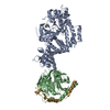
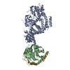
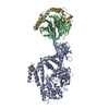
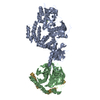
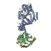
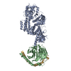
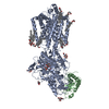
| Title | Human GRK2 in complex with human G-beta-gamma subunits and CCG211998 (14ak) | |||||||||||||||||||||||||||||||||||||||
 Components Components |
| |||||||||||||||||||||||||||||||||||||||
 Keywords Keywords | TRANSFERASE/TRANSFERASE INHIBITOR / kinase / inhibitor complex / TRANSFERASE-TRANSFERASE INHIBITOR complex | |||||||||||||||||||||||||||||||||||||||
| Function / homology |  Function and homology information Function and homology informationnegative regulation of the force of heart contraction by chemical signal / negative regulation of relaxation of smooth muscle / beta-adrenergic-receptor kinase / Edg-2 lysophosphatidic acid receptor binding / alpha-2A adrenergic receptor binding / beta-adrenergic receptor kinase activity / G protein-coupled receptor kinase activity / tachykinin receptor signaling pathway / negative regulation of striated muscle contraction / positive regulation of catecholamine secretion ...negative regulation of the force of heart contraction by chemical signal / negative regulation of relaxation of smooth muscle / beta-adrenergic-receptor kinase / Edg-2 lysophosphatidic acid receptor binding / alpha-2A adrenergic receptor binding / beta-adrenergic receptor kinase activity / G protein-coupled receptor kinase activity / tachykinin receptor signaling pathway / negative regulation of striated muscle contraction / positive regulation of catecholamine secretion / Activation of SMO / cytoplasmic side of mitochondrial outer membrane / regulation of the force of heart contraction / desensitization of G protein-coupled receptor signaling pathway / Calmodulin induced events / cardiac muscle contraction / viral genome replication / receptor internalization / G protein-coupled receptor binding / Olfactory Signaling Pathway / Activation of the phototransduction cascade / G protein-coupled acetylcholine receptor signaling pathway / G beta:gamma signalling through PLC beta / Presynaptic function of Kainate receptors / Thromboxane signalling through TP receptor / Activation of G protein gated Potassium channels / Inhibition of voltage gated Ca2+ channels via Gbeta/gamma subunits / G-protein activation / Glucagon signaling in metabolic regulation / Prostacyclin signalling through prostacyclin receptor / G beta:gamma signalling through CDC42 / Synthesis, secretion, and inactivation of Glucagon-like Peptide-1 (GLP-1) / G beta:gamma signalling through BTK / photoreceptor disc membrane / ADP signalling through P2Y purinoceptor 12 / Glucagon-type ligand receptors / Sensory perception of sweet, bitter, and umami (glutamate) taste / Adrenaline,noradrenaline inhibits insulin secretion / Vasopressin regulates renal water homeostasis via Aquaporins / Glucagon-like Peptide-1 (GLP1) regulates insulin secretion / G alpha (z) signalling events / cellular response to catecholamine stimulus / ADP signalling through P2Y purinoceptor 1 / ADORA2B mediated anti-inflammatory cytokines production / G beta:gamma signalling through PI3Kgamma / Cargo recognition for clathrin-mediated endocytosis / adenylate cyclase-activating dopamine receptor signaling pathway / Cooperation of PDCL (PhLP1) and TRiC/CCT in G-protein beta folding / GPER1 signaling / cellular response to prostaglandin E stimulus / heterotrimeric G-protein complex / G alpha (12/13) signalling events / Inactivation, recovery and regulation of the phototransduction cascade / G-protein beta-subunit binding / extracellular vesicle / sensory perception of taste / heart development / Thrombin signalling through proteinase activated receptors (PARs) / signaling receptor complex adaptor activity / presynapse / retina development in camera-type eye / GTPase binding / fibroblast proliferation / Ca2+ pathway / High laminar flow shear stress activates signaling by PIEZO1 and PECAM1:CDH5:KDR in endothelial cells / G alpha (i) signalling events / G alpha (s) signalling events / phospholipase C-activating G protein-coupled receptor signaling pathway / G alpha (q) signalling events / Ras protein signal transduction / protein kinase activity / Extra-nuclear estrogen signaling / cell population proliferation / postsynapse / cilium / G protein-coupled receptor signaling pathway / lysosomal membrane / GTPase activity / symbiont entry into host cell / synapse / protein-containing complex binding / signal transduction / extracellular exosome / ATP binding / membrane / plasma membrane / cytoplasm / cytosol Similarity search - Function | |||||||||||||||||||||||||||||||||||||||
| Biological species |  Homo sapiens (human) Homo sapiens (human) | |||||||||||||||||||||||||||||||||||||||
| Method |  X-RAY DIFFRACTION / SYNCHROTRON / MOLECULAR REPLACEMENT / Resolution: 2.6 Å X-RAY DIFFRACTION / SYNCHROTRON / MOLECULAR REPLACEMENT / Resolution: 2.6 Å | |||||||||||||||||||||||||||||||||||||||
 Authors Authors | Cato, M.C. / Homan, K.T. / Tesmer, J.J.G. | |||||||||||||||||||||||||||||||||||||||
| Funding support |  United States, 12items United States, 12items
| |||||||||||||||||||||||||||||||||||||||
 Citation Citation | Journal: J. Med. Chem. / Year: 2017 Title: Structure-Based Design of Highly Selective and Potent G Protein-Coupled Receptor Kinase 2 Inhibitors Based on Paroxetine. Authors: Waldschmidt, H.V. / Homan, K.T. / Cato, M.C. / Cruz-Rodriguez, O. / Cannavo, A. / Wilson, M.W. / Song, J. / Cheung, J.Y. / Koch, W.J. / Tesmer, J.J. / Larsen, S.D. | |||||||||||||||||||||||||||||||||||||||
| History |
|
- Structure visualization
| Structure viewer | Molecule: MolmilJmol/JSmol |
|---|
- Downloads & links
-Download
| PDBx/mmCIF format | 5ukk.cif.gz | 419.5 KB | Display | PDBx/mmCIF format |
|---|---|---|---|---|
| PDB format | pdb5ukk.ent.gz | 344.4 KB | Display | PDB format |
| PDBx/mmJSON format | 5ukk.json.gz | Tree view | PDBx/mmJSON format | |
| Others |  Other downloads Other downloads |
-Validation report
| Arichive directory | https://data.pdbj.org/pub/pdb/validation_reports/uk/5ukkftp://data.pdbj.org/pub/pdb/validation_reports/uk/5ukk | HTTPS FTP |
|---|
-Related structure data
| Related structure data | 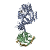 5uklC  5ukmC  3v5wS C: citing same article ( S: Starting model for refinement |
|---|---|
| Similar structure data |

-Links
 PDBj
PDBj


- Assembly
| Deposited unit | 
| ||||||||
|---|---|---|---|---|---|---|---|---|---|
| 1 |
| ||||||||
| Unit cell |
|
-Components
-Protein , 1 types, 1 molecules A
| #1: Protein | Mass: 74799.953 Da / Num. of mol.: 1 / Mutation: S670A Source method: isolated from a genetically manipulated source Source: (gene. exp.) Homo sapiens (human) / Gene: GRK2, ADRBK1, BARK, BARK1 / Plasmid: pFastBac / Production host:  Trichoplusia ni (cabbage looper) Trichoplusia ni (cabbage looper)References: UniProt: P25098, beta-adrenergic-receptor kinase |
|---|
-Guanine nucleotide-binding protein ... , 2 types, 2 molecules BG
| #2: Protein | Mass: 37285.734 Da / Num. of mol.: 1 Source method: isolated from a genetically manipulated source Source: (gene. exp.) Homo sapiens (human) / Gene: GNB1 / Plasmid: pFastBacDual / Production host: Trichoplusia ni (cabbage looper) / References: UniProt: P62873 |
|---|---|
| #3: Protein | Mass: 6347.342 Da / Num. of mol.: 1 Source method: isolated from a genetically manipulated source Source: (gene. exp.) Homo sapiens (human) / Gene: GNG2 / Plasmid: pFastBacDual / Production host: Trichoplusia ni (cabbage looper) / References: UniProt: P59768 |
-Non-polymers , 3 types, 45 molecules 

| #4: Chemical | ChemComp-MG /  Mass: 24.305 Da / Num. of mol.: 1 / Source method: obtained synthetically / Formula: Mg Mass: 24.305 Da / Num. of mol.: 1 / Source method: obtained synthetically / Formula: Mg |
|---|---|
| #5: Chemical | ChemComp-8DJ /  Mass: 463.501 Da / Num. of mol.: 1 / Source method: obtained synthetically / Formula: C26H26FN3O4 Mass: 463.501 Da / Num. of mol.: 1 / Source method: obtained synthetically / Formula: C26H26FN3O4 |
| #6: Water | ChemComp-HOH / Mass: 18.015 Da / Num. of mol.: 43 / Source method: isolated from a natural source / Formula: H2O |
-Experimental details
-Experiment
| Experiment | Method: X-RAY DIFFRACTION / Number of used crystals: 1 |
|---|
- Sample preparation
| Crystal | Density Matthews: 3.12 Å3/Da / Density % sol: 60.6 % |
|---|---|
| Crystal grow | Temperature: 277 K / Method: vapor diffusion, hanging drop / pH: 6 Details: 50 mM MES, 8-16% PEG3350, 0.8-1.2 M sodium chloride |
-Data collection
| Diffraction | Mean temperature: 100 K | ||||||||||||||||||||||||||||||||||||||||||||||||||||||||||||||||||||||||||||||||||||||||||||||||||||||||||||||||||||||||||||||
|---|---|---|---|---|---|---|---|---|---|---|---|---|---|---|---|---|---|---|---|---|---|---|---|---|---|---|---|---|---|---|---|---|---|---|---|---|---|---|---|---|---|---|---|---|---|---|---|---|---|---|---|---|---|---|---|---|---|---|---|---|---|---|---|---|---|---|---|---|---|---|---|---|---|---|---|---|---|---|---|---|---|---|---|---|---|---|---|---|---|---|---|---|---|---|---|---|---|---|---|---|---|---|---|---|---|---|---|---|---|---|---|---|---|---|---|---|---|---|---|---|---|---|---|---|---|---|---|
| Diffraction source | Source: SYNCHROTRON / Site: APS / Beamline: 21-ID-G / Wavelength: 0.97849 Å | ||||||||||||||||||||||||||||||||||||||||||||||||||||||||||||||||||||||||||||||||||||||||||||||||||||||||||||||||||||||||||||||
| Detector | Type: MARMOSAIC 300 mm CCD / Detector: CCD / Date: Jul 12, 2013 | ||||||||||||||||||||||||||||||||||||||||||||||||||||||||||||||||||||||||||||||||||||||||||||||||||||||||||||||||||||||||||||||
| Radiation | Monochromator: diamond(111) / Protocol: SINGLE WAVELENGTH / Monochromatic (M) / Laue (L): M / Scattering type: x-ray | ||||||||||||||||||||||||||||||||||||||||||||||||||||||||||||||||||||||||||||||||||||||||||||||||||||||||||||||||||||||||||||||
| Radiation wavelength | Wavelength: 0.97849 Å / Relative weight: 1 | ||||||||||||||||||||||||||||||||||||||||||||||||||||||||||||||||||||||||||||||||||||||||||||||||||||||||||||||||||||||||||||||
| Reflection | Resolution: 2.6→50 Å / Num. obs: 39963 / % possible obs: 92.7 % / Redundancy: 2.6 % / Rmerge(I) obs: 0.098 / Net I/σ(I): 9.24 | ||||||||||||||||||||||||||||||||||||||||||||||||||||||||||||||||||||||||||||||||||||||||||||||||||||||||||||||||||||||||||||||
| Reflection shell |
|
- Processing
| Software |
| ||||||||||||||||||||||||||||||||||||||||||||||||||||||||||||||||||||||||||||||||||||||||||||||||||||||||||||||||||||||||||||||||||||||||||||||||||||||
|---|---|---|---|---|---|---|---|---|---|---|---|---|---|---|---|---|---|---|---|---|---|---|---|---|---|---|---|---|---|---|---|---|---|---|---|---|---|---|---|---|---|---|---|---|---|---|---|---|---|---|---|---|---|---|---|---|---|---|---|---|---|---|---|---|---|---|---|---|---|---|---|---|---|---|---|---|---|---|---|---|---|---|---|---|---|---|---|---|---|---|---|---|---|---|---|---|---|---|---|---|---|---|---|---|---|---|---|---|---|---|---|---|---|---|---|---|---|---|---|---|---|---|---|---|---|---|---|---|---|---|---|---|---|---|---|---|---|---|---|---|---|---|---|---|---|---|---|---|---|---|---|
| Refinement | Method to determine structure: MOLECULAR REPLACEMENT Starting model: PDB entry 3V5W Resolution: 2.6→30 Å / Cor.coef. Fo:Fc: 0.946 / Cor.coef. Fo:Fc free: 0.911 / SU B: 39.783 / SU ML: 0.36 / SU R Cruickshank DPI: 0.9635 / Cross valid method: THROUGHOUT / σ(F): 0 / ESU R: 0.963 / ESU R Free: 0.372 / Details: HYDROGENS HAVE BEEN ADDED IN THE RIDING POSITIONS
| ||||||||||||||||||||||||||||||||||||||||||||||||||||||||||||||||||||||||||||||||||||||||||||||||||||||||||||||||||||||||||||||||||||||||||||||||||||||
| Solvent computation | Ion probe radii: 0.8 Å / Shrinkage radii: 0.8 Å / VDW probe radii: 1.2 Å | ||||||||||||||||||||||||||||||||||||||||||||||||||||||||||||||||||||||||||||||||||||||||||||||||||||||||||||||||||||||||||||||||||||||||||||||||||||||
| Displacement parameters | Biso max: 194.75 Å2 / Biso mean: 79.624 Å2 / Biso min: 31.95 Å2
| ||||||||||||||||||||||||||||||||||||||||||||||||||||||||||||||||||||||||||||||||||||||||||||||||||||||||||||||||||||||||||||||||||||||||||||||||||||||
| Refinement step | Cycle: final / Resolution: 2.6→30 Å
| ||||||||||||||||||||||||||||||||||||||||||||||||||||||||||||||||||||||||||||||||||||||||||||||||||||||||||||||||||||||||||||||||||||||||||||||||||||||
| Refine LS restraints |
| ||||||||||||||||||||||||||||||||||||||||||||||||||||||||||||||||||||||||||||||||||||||||||||||||||||||||||||||||||||||||||||||||||||||||||||||||||||||
| LS refinement shell | Resolution: 2.6→2.667 Å / Rfactor Rfree error: 0 / Total num. of bins used: 20
| ||||||||||||||||||||||||||||||||||||||||||||||||||||||||||||||||||||||||||||||||||||||||||||||||||||||||||||||||||||||||||||||||||||||||||||||||||||||
| Refinement TLS params. | Method: refined / Refine-ID: X-RAY DIFFRACTION
| ||||||||||||||||||||||||||||||||||||||||||||||||||||||||||||||||||||||||||||||||||||||||||||||||||||||||||||||||||||||||||||||||||||||||||||||||||||||
| Refinement TLS group |
|
