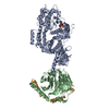
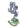
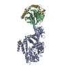
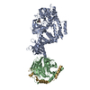
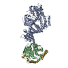
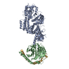
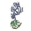
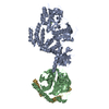
[English] 日本語
Yorodumi
- PDB-3v5w: Human G Protein-Coupled Receptor Kinase 2 in Complex with Soluble... -

+
Open data


ID or keywords:

Loading...

-
Basic information

Entry
Database: PDB / ID: 3v5w
TitleHuman G Protein-Coupled Receptor Kinase 2 in Complex with Soluble Gbetagamma Subunits and Paroxetine
Components
  • (Guanine nucleotide-binding protein ...) x 2
  • G-protein coupled receptor kinase 2
KeywordsTRANSFERASE/TRANSFERASE INHIBITOR / Inhibitor complex / Protein kinase / Beta propeller / RGS homology domain / Pleckstrin homology domain / Kinase / Signal transduction / Peripheral membrane protein / TRANSFERASE-TRANSFERASE INHIBITOR complex
Function / homology
Function and homology information


beta-adrenergic-receptor kinase / negative regulation of the force of heart contraction by chemical signal / negative regulation of relaxation of smooth muscle / beta-adrenergic receptor kinase activity / G protein-coupled receptor kinase activity / Edg-2 lysophosphatidic acid receptor binding / alpha-2A adrenergic receptor binding / positive regulation of catecholamine secretion / tachykinin receptor signaling pathway / Olfactory Signaling Pathway ...beta-adrenergic-receptor kinase / negative regulation of the force of heart contraction by chemical signal / negative regulation of relaxation of smooth muscle / beta-adrenergic receptor kinase activity / G protein-coupled receptor kinase activity / Edg-2 lysophosphatidic acid receptor binding / alpha-2A adrenergic receptor binding / positive regulation of catecholamine secretion / tachykinin receptor signaling pathway / Olfactory Signaling Pathway / Sensory perception of sweet, bitter, and umami (glutamate) taste / Synthesis, secretion, and inactivation of Glucagon-like Peptide-1 (GLP-1) / Activation of SMO / negative regulation of striated muscle contraction / desensitization of G protein-coupled receptor signaling pathway / regulation of the force of heart contraction / Activation of the phototransduction cascade / Activation of G protein gated Potassium channels / G-protein activation / G beta:gamma signalling through PI3Kgamma / Prostacyclin signalling through prostacyclin receptor / G beta:gamma signalling through PLC beta / ADP signalling through P2Y purinoceptor 1 / Thromboxane signalling through TP receptor / Presynaptic function of Kainate receptors / G beta:gamma signalling through CDC42 / Inhibition of voltage gated Ca2+ channels via Gbeta/gamma subunits / Glucagon-type ligand receptors / Adrenaline,noradrenaline inhibits insulin secretion / G alpha (12/13) signalling events / G beta:gamma signalling through BTK / ADP signalling through P2Y purinoceptor 12 / Cooperation of PDCL (PhLP1) and TRiC/CCT in G-protein beta folding / Thrombin signalling through proteinase activated receptors (PARs) / Ca2+ pathway / G alpha (z) signalling events / Extra-nuclear estrogen signaling / G alpha (s) signalling events / Calmodulin induced events / G alpha (q) signalling events / G alpha (i) signalling events / Glucagon-like Peptide-1 (GLP1) regulates insulin secretion / Vasopressin regulates renal water homeostasis via Aquaporins / cardiac muscle contraction / viral genome replication / G protein-coupled receptor binding / G protein-coupled acetylcholine receptor signaling pathway / cilium / receptor internalization / photoreceptor disc membrane / cellular response to catecholamine stimulus / adenylate cyclase-activating dopamine receptor signaling pathway / cellular response to prostaglandin E stimulus / sensory perception of taste / G-protein beta-subunit binding / heterotrimeric G-protein complex / signaling receptor complex adaptor activity / Cargo recognition for clathrin-mediated endocytosis / presynapse / retina development in camera-type eye / GTPase binding / phospholipase C-activating G protein-coupled receptor signaling pathway / heart development / G alpha (s) signalling events / postsynapse / G alpha (q) signalling events / peptidyl-serine phosphorylation / cell population proliferation / protein kinase activity / symbiont entry into host cell / G protein-coupled receptor signaling pathway / GTPase activity / protein-containing complex binding / ATP binding / membrane / plasma membrane / cytosol / cytoplasm
Similarity search - Function
Helix Hairpins - #1270 / GPCR kinase / Transducin (heterotrimeric G protein), gamma chain / G Protein Gi Gamma 2 / Regulator of G-protein Signalling 4, domain 2 / Regulator of G-protein Signalling 4; domain 2 / Regulator of G protein signaling domain / RGS, subdomain 2 / RGS domain / RGS domain profile. ...Helix Hairpins - #1270 / GPCR kinase / Transducin (heterotrimeric G protein), gamma chain / G Protein Gi Gamma 2 / Regulator of G-protein Signalling 4, domain 2 / Regulator of G-protein Signalling 4; domain 2 / Regulator of G protein signaling domain / RGS, subdomain 2 / RGS domain / RGS domain profile. / Regulator of G protein signalling domain / RGS domain superfamily / Pleckstrin-homology domain (PH domain)/Phosphotyrosine-binding domain (PTB) / PH-domain like / YVTN repeat-like/Quinoprotein amine dehydrogenase / Extension to Ser/Thr-type protein kinases / AGC-kinase, C-terminal / AGC-kinase C-terminal domain profile. / 7 Propeller / Methylamine Dehydrogenase; Chain H / PH domain / PH domain profile. / Pleckstrin homology domain. / Pleckstrin homology domain / G-protein, gamma subunit / G-protein gamma subunit domain profile. / GGL domain / G-protein gamma-like domain superfamily / G-protein gamma-like domain / GGL domain / G protein gamma subunit-like motifs / Guanine nucleotide-binding protein, beta subunit / G-protein, beta subunit / Few Secondary Structures / Irregular / Helix Hairpins / PH-like domain superfamily / G-protein beta WD-40 repeat / WD40 repeat, conserved site / Trp-Asp (WD) repeats signature. / Roll / WD domain, G-beta repeat / WD40 repeats / WD40 repeat / Trp-Asp (WD) repeats profile. / Trp-Asp (WD) repeats circular profile. / WD40-repeat-containing domain superfamily / Transferase(Phosphotransferase) domain 1 / Transferase(Phosphotransferase); domain 1 / Phosphorylase Kinase; domain 1 / Phosphorylase Kinase; domain 1 / Serine/threonine-protein kinase, active site / Serine/Threonine protein kinases active-site signature. / WD40/YVTN repeat-like-containing domain superfamily / Protein kinase domain / Serine/Threonine protein kinases, catalytic domain / Protein kinase, ATP binding site / Protein kinases ATP-binding region signature. / Protein kinase domain profile. / Protein kinase domain / Protein kinase-like domain superfamily / 2-Layer Sandwich / Orthogonal Bundle / Mainly Beta / Mainly Alpha / Alpha Beta
Similarity search - Domain/homology
Paroxetine / Beta-adrenergic receptor kinase 1 / Guanine nucleotide-binding protein G(I)/G(S)/G(T) subunit beta-1 / Guanine nucleotide-binding protein G(I)/G(S)/G(O) subunit gamma-2
Similarity search - Component
Biological speciesHomo sapiens (human)
Bos taurus (cattle)
MethodX-RAY DIFFRACTION / SYNCHROTRON / MOLECULAR REPLACEMENT / Resolution: 2.07 Å
AuthorsThal, D.M. / Tesmer, J.J.G.
CitationJournal: Acs Chem.Biol. / Year: 2012
Title: Paroxetine is a direct inhibitor of g protein-coupled receptor kinase 2 and increases myocardial contractility.
Authors: Thal, D.M. / Homan, K.T. / Chen, J. / Wu, E.K. / Hinkle, P.M. / Huang, Z.M. / Chuprun, J.K. / Song, J. / Gao, E. / Cheung, J.Y. / Sklar, L.A. / Koch, W.J. / Tesmer, J.J.
History
DepositionDec 17, 2011Deposition site: RCSB / Processing site: RCSB
Revision 1.0Sep 26, 2012Provider: repository / Type: Initial release
Revision 1.1Dec 12, 2012Group: Database references
Revision 1.2Sep 13, 2023Group: Data collection / Database references ...Data collection / Database references / Derived calculations / Refinement description
Category: chem_comp_atom / chem_comp_bond ...chem_comp_atom / chem_comp_bond / database_2 / pdbx_initial_refinement_model / pdbx_struct_conn_angle / struct_conn / struct_ref_seq_dif / struct_site
Item: _database_2.pdbx_DOI / _database_2.pdbx_database_accession ..._database_2.pdbx_DOI / _database_2.pdbx_database_accession / _pdbx_struct_conn_angle.ptnr1_auth_comp_id / _pdbx_struct_conn_angle.ptnr1_auth_seq_id / _pdbx_struct_conn_angle.ptnr1_label_comp_id / _pdbx_struct_conn_angle.ptnr1_label_seq_id / _pdbx_struct_conn_angle.ptnr3_auth_comp_id / _pdbx_struct_conn_angle.ptnr3_auth_seq_id / _pdbx_struct_conn_angle.ptnr3_label_comp_id / _pdbx_struct_conn_angle.ptnr3_label_seq_id / _pdbx_struct_conn_angle.value / _struct_conn.pdbx_dist_value / _struct_conn.ptnr1_auth_comp_id / _struct_conn.ptnr1_auth_seq_id / _struct_conn.ptnr1_label_comp_id / _struct_conn.ptnr1_label_seq_id / _struct_ref_seq_dif.details / _struct_site.pdbx_auth_asym_id / _struct_site.pdbx_auth_comp_id / _struct_site.pdbx_auth_seq_id

-
Structure visualization

Structure viewerMolecule:
MolmilJmol/JSmol

Downloads & links

-
Assembly

Deposited unit
A: G-protein coupled receptor kinase 2
B: Guanine nucleotide-binding protein G(I)/G(S)/G(T) subunit beta-1
G: Guanine nucleotide-binding protein G(I)/G(S)/G(O) subunit gamma-2
hetero molecules


Theoretical massNumber of molelcules
Total (without water)126,1375
Polymers125,7843
Non-polymers3542
Water5,332296
1


  • Idetical with deposited unit
  • defined by author&software
TypeNameSymmetry operationNumber
identity operation1_555x,y,z1
Buried area6540 Å2
ΔGint-49 kcal/mol
Surface area46190 Å2
MethodPISA
Unit cell
Length a, b, c (Å)194.973, 71.011, 111.169
Angle α, β, γ (deg.)90.00, 110.41, 90.00
Int Tables number5
Space group name H-MC121
Components on special symmetry positions
IDModelComponents
11A-833-

HOH

-
Components

-
Protein , 1 types, 1 molecules A

#1: Protein G-protein coupled receptor kinase 2 / Beta-ARK-1 / Beta-adrenergic receptor kinase 1


Mass: 79692.641 Da / Num. of mol.: 1
Source method: isolated from a genetically manipulated source
Source: (gene. exp.) Homo sapiens (human) / Gene: ADRBK1, BARK, BARK1, GRK2 / Production host: Spodoptera frugiperda (fall armyworm)
References: UniProt: P25098, beta-adrenergic-receptor kinase

-
Guanine nucleotide-binding protein ... , 2 types, 2 molecules BG

#2: Protein Guanine nucleotide-binding protein G(I)/G(S)/G(T) subunit beta-1 / Transducin beta chain 1


Mass: 37416.930 Da / Num. of mol.: 1
Source method: isolated from a genetically manipulated source
Source: (gene. exp.) Bos taurus (cattle) / Gene: GNB1 / Production host: Trichoplusia ni (cabbage looper) / References: UniProt: P62871
#3: Protein Guanine nucleotide-binding protein G(I)/G(S)/G(O) subunit gamma-2 / G gamma-I


Mass: 8673.959 Da / Num. of mol.: 1 / Mutation: C68S
Source method: isolated from a genetically manipulated source
Source: (gene. exp.) Bos taurus (cattle) / Gene: GNG2 / Production host: Trichoplusia ni (cabbage looper) / References: UniProt: P63212

-
Non-polymers , 3 types, 298 molecules

#4: Chemical ChemComp-8PR / Paroxetine / (3S,4R)-3-[(1,3-benzodioxol-5-yloxy)methyl]-4-(4-fluorophenyl)piperidine / Paroxetine


Mass: 329.365 Da / Num. of mol.: 1 / Source method: obtained synthetically / Formula: C19H20FNO3 / Comment: antidepressant, inhibitor*YM
#5: Chemical ChemComp-MG / MAGNESIUM ION


Mass: 24.305 Da / Num. of mol.: 1 / Source method: obtained synthetically / Formula: Mg
#6: Water ChemComp-HOH / water / Water


Mass: 18.015 Da / Num. of mol.: 296 / Source method: isolated from a natural source / Formula: H2O

-
Experimental details

-
Experiment

ExperimentMethod: X-RAY DIFFRACTION / Number of used crystals: 1

-
Sample preparation

CrystalDensity Matthews: 2.87 Å3/Da / Density % sol: 57.1 %
Crystal growTemperature: 277 K / Method: vapor diffusion, hanging drop / pH: 5.8
Details: 100 mM MES pH 5.8, 200 mM NaCl, and 8% (w/v) PEG3350, VAPOR DIFFUSION, HANGING DROP, temperature 277K

-
Data collection

DiffractionMean temperature: 110 K
Diffraction sourceSource: SYNCHROTRON / Site: APS / Beamline: 21-ID-F / Wavelength: 0.97857 Å
DetectorType: RAYONIX MX-225 / Detector: CCD / Date: Mar 21, 2011 / Details: Be Lenses/Diamond Laue Mono
RadiationMonochromator: Diamond [111] / Protocol: SINGLE WAVELENGTH / Monochromatic (M) / Laue (L): M / Scattering type: x-ray
Radiation wavelengthWavelength: 0.97857 Å / Relative weight: 1
ReflectionResolution: 2.07→25 Å / Num. all: 55040 / Num. obs: 55040 / % possible obs: 61.8 % / Observed criterion σ(F): -99999999 / Observed criterion σ(I): -3 / Redundancy: 2.6 % / Rsym value: 0.077 / Net I/σ(I): 16.8
Reflection shellResolution: 2.07→2.11 Å / Redundancy: 1.2 % / Mean I/σ(I) obs: 1.5 / Num. unique all: 12 / Rsym value: 0.597 / % possible all: 30

-
Processing

Software
NameVersionClassification
MD2diffractometer software from EMBL (with LS-CAT developed extensions)data collection
PHASERphasing
REFMAC5.6.0117refinement
HKL-2000data reduction
SCALEPACKdata scaling
RefinementMethod to determine structure: MOLECULAR REPLACEMENT
Starting model: 1OMW
Resolution: 2.07→24.53 Å / Cor.coef. Fo:Fc: 0.957 / Cor.coef. Fo:Fc free: 0.927 / SU B: 13.518 / SU ML: 0.149 / Isotropic thermal model: Individual isotropic / Cross valid method: THROUGHOUT / σ(F): -9999999 / ESU R: 0.324 / ESU R Free: 0.244 / Stereochemistry target values: MAXIMUM LIKELIHOOD / Details: HYDROGENS HAVE BEEN ADDED IN THE RIDING POSITIONS
RfactorNum. reflection% reflectionSelection details
Rfree0.24668 2764 5 %RANDOM
Rwork0.19349 ---
all0.19614 52208 --
obs0.19614 52208 62.9 %-
Solvent computationIon probe radii: 0.8 Å / Shrinkage radii: 0.8 Å / VDW probe radii: 1.2 Å / Solvent model: MASK
Displacement parametersBiso mean: 45.265 Å2
Baniso -1Baniso -2Baniso -3
1--0.24 Å20 Å2-0.66 Å2
2--0.2 Å20 Å2
3----0.43 Å2
Refinement stepCycle: LAST / Resolution: 2.07→24.53 Å
ProteinNucleic acidLigandSolventTotal
Num. atoms8152 0 25 296 8473
Refine LS restraints
Refine-IDTypeDev idealDev ideal targetNumber
X-RAY DIFFRACTIONr_bond_refined_d0.0080.0198360
X-RAY DIFFRACTIONr_bond_other_d0.0010.025846
X-RAY DIFFRACTIONr_angle_refined_deg1.2011.9611269
X-RAY DIFFRACTIONr_angle_other_deg0.81314179
X-RAY DIFFRACTIONr_dihedral_angle_1_deg5.95651018
X-RAY DIFFRACTIONr_dihedral_angle_2_deg32.93223.753405
X-RAY DIFFRACTIONr_dihedral_angle_3_deg15.67151513
X-RAY DIFFRACTIONr_dihedral_angle_4_deg15.8541566
X-RAY DIFFRACTIONr_chiral_restr0.0640.21212
X-RAY DIFFRACTIONr_gen_planes_refined0.0040.029280
X-RAY DIFFRACTIONr_gen_planes_other0.0010.021754
X-RAY DIFFRACTIONr_nbd_refined
X-RAY DIFFRACTIONr_nbd_other
X-RAY DIFFRACTIONr_nbtor_refined
X-RAY DIFFRACTIONr_nbtor_other
X-RAY DIFFRACTIONr_xyhbond_nbd_refined
X-RAY DIFFRACTIONr_xyhbond_nbd_other
X-RAY DIFFRACTIONr_metal_ion_refined
X-RAY DIFFRACTIONr_metal_ion_other
X-RAY DIFFRACTIONr_symmetry_vdw_refined
X-RAY DIFFRACTIONr_symmetry_vdw_other
X-RAY DIFFRACTIONr_symmetry_hbond_refined
X-RAY DIFFRACTIONr_symmetry_hbond_other
X-RAY DIFFRACTIONr_symmetry_metal_ion_refined
X-RAY DIFFRACTIONr_symmetry_metal_ion_other
X-RAY DIFFRACTIONr_mcbond_it
X-RAY DIFFRACTIONr_mcbond_other
X-RAY DIFFRACTIONr_mcangle_it
X-RAY DIFFRACTIONr_scbond_it
X-RAY DIFFRACTIONr_scangle_it
X-RAY DIFFRACTIONr_rigid_bond_restr
X-RAY DIFFRACTIONr_sphericity_free
X-RAY DIFFRACTIONr_sphericity_bonded
LS refinement shellResolution: 2.067→2.12 Å / Total num. of bins used: 20
RfactorNum. reflection% reflection
Rfree0.32 6 -
Rwork0.307 85 -
obs--1.42 %
Refinement TLS params.

Method: refined / Refine-ID: X-RAY DIFFRACTION

IDL112)L122)L132)L222)L232)L332)S11 (Å °)S12 (Å °)S13 (Å °)S21 (Å °)S22 (Å °)S23 (Å °)S31 (Å °)S32 (Å °)S33 (Å °)T112)T122)T132)T222)T232)T332)Origin x (Å)Origin y (Å)Origin z (Å)
10.63-0.2598-0.33030.14640.35811.61790.0389-0.05160.0377-0.0360.0111-0.0129-0.03980.0654-0.050.1501-0.0025-0.00980.08470.0010.0575-3.901920.654252.5371
20.4901-0.03940.71250.2646-0.59912.29220.46480.1595-0.1391-0.028-0.05230.16570.53490.3662-0.41250.48030.1322-0.19020.0663-0.06840.19458.0086-12.966158.517
30.3339-0.19650.66340.2849-0.45292.09320.07690.05420.0719-0.0331-0.06420.05660.33940.2247-0.01280.14250.09470.00780.1450.00010.0966-5.875915.262416.3332
40.7276-0.1650.14020.2636-0.16540.56-0.0207-0.01530.0271-0.0130.03880.03470.0124-0.0021-0.01810.0750.00480.00910.09720.0220.1116-28.612934.0724-3.1479
Refinement TLS group
IDRefine-IDRefine TLS-IDAuth asym-IDAuth seq-ID
1X-RAY DIFFRACTION1A30 - 184
2X-RAY DIFFRACTION1A513 - 545
3X-RAY DIFFRACTION1A801 - 882
4X-RAY DIFFRACTION2A185 - 512
5X-RAY DIFFRACTION2A701 - 702
6X-RAY DIFFRACTION2A883 - 925
7X-RAY DIFFRACTION3A546 - 668
8X-RAY DIFFRACTION3A926 - 956
9X-RAY DIFFRACTION4B2 - 340
10X-RAY DIFFRACTION4G8 - 64
11X-RAY DIFFRACTION4B401 - 540

+
About Yorodumi

-
News

-
Feb 9, 2022. New format data for meta-information of EMDB entries

New format data for meta-information of EMDB entries

  • Version 3 of the EMDB header file is now the official format.
  • The previous official version 1.9 will be removed from the archive.

Related info.:EMDB header

External links:wwPDB to switch to version 3 of the EMDB data model

-
Aug 12, 2020. Covid-19 info

Covid-19 info

URL: https://pdbj.org/emnavi/covid19.php

New page: Covid-19 featured information page in EM Navigator.

Related info.:Covid-19 info / Mar 5, 2020. Novel coronavirus structure data

+
Mar 5, 2020. Novel coronavirus structure data

Novel coronavirus structure data

Related info.:Yorodumi Speices / Aug 12, 2020. Covid-19 info

External links:COVID-19 featured content - PDBj / Molecule of the Month (242):Coronavirus Proteases

+
Jan 31, 2019. EMDB accession codes are about to change! (news from PDBe EMDB page)

EMDB accession codes are about to change! (news from PDBe EMDB page)

  • The allocation of 4 digits for EMDB accession codes will soon come to an end. Whilst these codes will remain in use, new EMDB accession codes will include an additional digit and will expand incrementally as the available range of codes is exhausted. The current 4-digit format prefixed with “EMD-” (i.e. EMD-XXXX) will advance to a 5-digit format (i.e. EMD-XXXXX), and so on. It is currently estimated that the 4-digit codes will be depleted around Spring 2019, at which point the 5-digit format will come into force.
  • The EM Navigator/Yorodumi systems omit the EMD- prefix.

Related info.:Q: What is EMD? / ID/Accession-code notation in Yorodumi/EM Navigator

External links:EMDB Accession Codes are Changing Soon! / Contact to PDBj

+
Jul 12, 2017. Major update of PDB

Major update of PDB

  • wwPDB released updated PDB data conforming to the new PDBx/mmCIF dictionary.
  • This is a major update changing the version number from 4 to 5, and with Remediation, in which all the entries are updated.
  • In this update, many items about electron microscopy experimental information are reorganized (e.g. em_software).
  • Now, EM Navigator and Yorodumi are based on the updated data.

External links:wwPDB Remediation / Enriched Model Files Conforming to OneDep Data Standards Now Available in the PDB FTP Archive

-
Yorodumi

Thousand views of thousand structures

  • Yorodumi is a browser for structure data from EMDB, PDB, SASBDB, etc.
  • This page is also the successor to EM Navigator detail page, and also detail information page/front-end page for Omokage search.
  • The word "yorodu" (or yorozu) is an old Japanese word meaning "ten thousand". "mi" (miru) is to see.

Related info.:EMDB / PDB / SASBDB / Comparison of 3 databanks / Yorodumi Search / Aug 31, 2016. New EM Navigator & Yorodumi / Yorodumi Papers / Jmol/JSmol / Function and homology information / Changes in new EM Navigator and Yorodumi

Read more