 Movie
Movie Controller

[English] 日本語
 Yorodumi
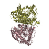
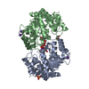
- PDB-4j7a: Crystal Structure of Est25 - a Bacterial Homolog of Hormone-Sensi... -
+ Open data

- Basic information
| Entry | Database: PDB / ID: 4j7a | ||||||
|---|---|---|---|---|---|---|---|
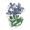
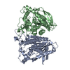
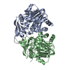
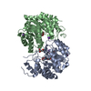
| Title | Crystal Structure of Est25 - a Bacterial Homolog of Hormone-Sensitive Lipase from a Metagenomic Library | ||||||
 Components Components | Esterase | ||||||
 Keywords Keywords | HYDROLASE / alpha/beta | ||||||
| Function / homology |  Function and homology information Function and homology information | ||||||
| Biological species |  uncultured bacterium (environmental samples) uncultured bacterium (environmental samples) | ||||||
| Method |  X-RAY DIFFRACTION / SYNCHROTRON / MAD / Resolution: 1.492 Å X-RAY DIFFRACTION / SYNCHROTRON / MAD / Resolution: 1.492 Å | ||||||
 Authors Authors | Ngo, T.D. / Ryu, B.H. / Ju, H.S. / Jang, E.J. / Park, K.S. / Joo, S.B. / Kim, K.K. / Kim, D.H. | ||||||
 Citation Citation | Journal: Acta Crystallogr.,Sect.D / Year: 2014 Title: Structural and functional analyses of a bacterial homologue of hormone-sensitive lipase from a metagenomic library Authors: Ngo, T.D. / Ryu, B.H. / Ju, H. / Jang, E. / Park, K. / Kim, K.K. / Kim, D.H. | ||||||
| History |
|
- Structure visualization
| Structure viewer | Molecule: MolmilJmol/JSmol |
|---|
- Downloads & links
-Download
| PDBx/mmCIF format | 4j7a.cif.gz | 564.6 KB | Display | PDBx/mmCIF format |
|---|---|---|---|---|
| PDB format | pdb4j7a.ent.gz | 465.4 KB | Display | PDB format |
| PDBx/mmJSON format | 4j7a.json.gz | Tree view | PDBx/mmJSON format | |
| Others |  Other downloads Other downloads |
-Validation report
| Arichive directory | https://data.pdbj.org/pub/pdb/validation_reports/j7/4j7aftp://data.pdbj.org/pub/pdb/validation_reports/j7/4j7a | HTTPS FTP |
|---|
-Related structure data
| Similar structure data |
|---|


-Links
 PDBj
PDBj- Assembly
| Deposited unit | 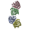
| |||||||||
|---|---|---|---|---|---|---|---|---|---|---|
| 1 | 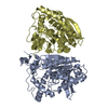
| |||||||||
| 2 | 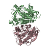
| |||||||||
| Unit cell |
| |||||||||
| Components on special symmetry positions |
|
-Components
| #1: Protein | Mass: 39744.430 Da / Num. of mol.: 4 Source method: isolated from a genetically manipulated source Source: (gene. exp.) uncultured bacterium (environmental samples)Plasmid: pQE30 / Production host: #2: Water | ChemComp-HOH / |  Mass: 18.015 Da / Num. of mol.: 2111 / Source method: isolated from a natural source / Formula: H2O Mass: 18.015 Da / Num. of mol.: 2111 / Source method: isolated from a natural source / Formula: H2O |
|---|
-Experimental details
-Experiment
| Experiment | Method: X-RAY DIFFRACTION / Number of used crystals: 1 |
|---|
- Sample preparation
| Crystal | Density Matthews: 2.92 Å3/Da / Density % sol: 57.9 % |
|---|---|
| Crystal grow | Temperature: 295 K / Method: microbatch / pH: 7 Details: 2.4M sodium malonate pH 7.0, Microbatch, temperature 295K |
-Data collection
| Diffraction | Mean temperature: 203 K | |||||||||||||||
|---|---|---|---|---|---|---|---|---|---|---|---|---|---|---|---|---|
| Diffraction source | Source: SYNCHROTRON / Site: Photon Factory  / Beamline: AR-NW12A / Wavelength: 0.97888, 0.97917, 0.96395, 1.00000 / Beamline: AR-NW12A / Wavelength: 0.97888, 0.97917, 0.96395, 1.00000 | |||||||||||||||
| Detector | Type: ADSC QUANTUM 210 / Detector: CCD | |||||||||||||||
| Radiation | Monochromator: GRAPHITE / Protocol: MAD / Monochromatic (M) / Laue (L): M / Scattering type: x-ray | |||||||||||||||
| Radiation wavelength |
| |||||||||||||||
| Reflection | Resolution: 1.49→50 Å / Num. all: 297211 / Num. obs: 285917 / % possible obs: 96.2 % / Observed criterion σ(F): 3 / Observed criterion σ(I): 3 | |||||||||||||||
| Reflection shell | Resolution: 1.49→50 Å / Redundancy: 3.3 % / Rmerge(I) obs: 0.316 / Mean I/σ(I) obs: 3.6 / % possible all: 96.2 |
- Processing
| Software |
| ||||||||||||||||||||||||||||||||||||||||||||||||||||||||||||||||||||||||||||||||||||||||||||||||||||||||||||||||||||||||||||||||||||||||||||||||||||||||||||||||||||||||||||||||||||||||||
|---|---|---|---|---|---|---|---|---|---|---|---|---|---|---|---|---|---|---|---|---|---|---|---|---|---|---|---|---|---|---|---|---|---|---|---|---|---|---|---|---|---|---|---|---|---|---|---|---|---|---|---|---|---|---|---|---|---|---|---|---|---|---|---|---|---|---|---|---|---|---|---|---|---|---|---|---|---|---|---|---|---|---|---|---|---|---|---|---|---|---|---|---|---|---|---|---|---|---|---|---|---|---|---|---|---|---|---|---|---|---|---|---|---|---|---|---|---|---|---|---|---|---|---|---|---|---|---|---|---|---|---|---|---|---|---|---|---|---|---|---|---|---|---|---|---|---|---|---|---|---|---|---|---|---|---|---|---|---|---|---|---|---|---|---|---|---|---|---|---|---|---|---|---|---|---|---|---|---|---|---|---|---|---|---|---|---|---|
| Refinement | Method to determine structure: MAD / Resolution: 1.492→28.555 Å / SU ML: 0.11 / σ(F): 1.34 / Phase error: 14.9 / Stereochemistry target values: ML
| ||||||||||||||||||||||||||||||||||||||||||||||||||||||||||||||||||||||||||||||||||||||||||||||||||||||||||||||||||||||||||||||||||||||||||||||||||||||||||||||||||||||||||||||||||||||||||
| Solvent computation | Shrinkage radii: 0.9 Å / VDW probe radii: 1.11 Å / Solvent model: FLAT BULK SOLVENT MODEL | ||||||||||||||||||||||||||||||||||||||||||||||||||||||||||||||||||||||||||||||||||||||||||||||||||||||||||||||||||||||||||||||||||||||||||||||||||||||||||||||||||||||||||||||||||||||||||
| Refinement step | Cycle: LAST / Resolution: 1.492→28.555 Å
| ||||||||||||||||||||||||||||||||||||||||||||||||||||||||||||||||||||||||||||||||||||||||||||||||||||||||||||||||||||||||||||||||||||||||||||||||||||||||||||||||||||||||||||||||||||||||||
| Refine LS restraints |
| ||||||||||||||||||||||||||||||||||||||||||||||||||||||||||||||||||||||||||||||||||||||||||||||||||||||||||||||||||||||||||||||||||||||||||||||||||||||||||||||||||||||||||||||||||||||||||
| LS refinement shell | Refine-ID: X-RAY DIFFRACTION / Total num. of bins used: 30
|
