 Movie
Movie Controller

[English] 日本語
 Yorodumi
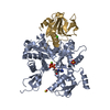
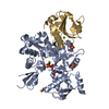
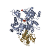
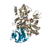
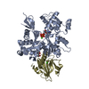
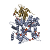
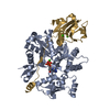
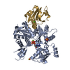
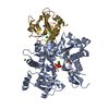
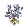
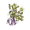
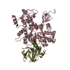
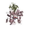
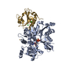
- PDB-1c0f: CRYSTAL STRUCTURE OF DICTYOSTELIUM CAATP-ACTIN IN COMPLEX WITH GE... -
+ Open data

- Basic information
| Entry | Database: PDB / ID: 1c0f | ||||||
|---|---|---|---|---|---|---|---|
| Title | CRYSTAL STRUCTURE OF DICTYOSTELIUM CAATP-ACTIN IN COMPLEX WITH GELSOLIN SEGMENT 1 | ||||||
 Components Components |
| ||||||
 Keywords Keywords | CONTRACTILE PROTEIN / CA ACTIN | ||||||
| Function / homology |  Function and homology information Function and homology informationintranuclear rod / leading edge of lamellipodium / phototaxis / phagolysosome / macropinocytic cup / striated muscle atrophy / regulation of establishment of T cell polarity / regulation of plasma membrane raft polarization / regulation of receptor clustering / positive regulation of keratinocyte apoptotic process ...intranuclear rod / leading edge of lamellipodium / phototaxis / phagolysosome / macropinocytic cup / striated muscle atrophy / regulation of establishment of T cell polarity / regulation of plasma membrane raft polarization / regulation of receptor clustering / positive regulation of keratinocyte apoptotic process / renal protein absorption / positive regulation of protein processing in phagocytic vesicle / cell pole / phosphatidylinositol 3-kinase catalytic subunit binding / positive regulation of actin nucleation / actin cap / myosin II binding / host-mediated suppression of symbiont invasion / actin filament severing / barbed-end actin filament capping / early phagosome / cell projection assembly / actin polymerization or depolymerization / actin filament depolymerization / plasma membrane repair / actin filament capping / hyperosmotic response / relaxation of cardiac muscle / Sensory processing of sound by outer hair cells of the cochlea / phagocytosis, engulfment / cardiac muscle cell contraction / cortical actin cytoskeleton / myosin binding / hepatocyte apoptotic process / cell leading edge / pseudopodium / mitotic cytokinesis / sarcoplasm / cilium assembly / Caspase-mediated cleavage of cytoskeletal proteins / endocytic vesicle / response to cAMP / phagocytosis / phagocytic cup / phagocytic vesicle / response to muscle stretch / vesicle-mediated transport / actin filament polymerization / phosphatidylinositol-4,5-bisphosphate binding / lipid droplet / actin filament organization / central nervous system development / actin filament / structural constituent of cytoskeleton / protein destabilization / cellular response to type II interferon / Hydrolases; Acting on acid anhydrides; Acting on acid anhydrides to facilitate cellular and subcellular movement / cell morphogenesis / chemotaxis / endocytosis / cell-cell junction / actin filament binding / lamellipodium / actin cytoskeleton / actin binding / secretory granule lumen / blood microparticle / cell cortex / amyloid fibril formation / ficolin-1-rich granule lumen / Amyloid fiber formation / focal adhesion / hydrolase activity / calcium ion binding / Neutrophil degranulation / positive regulation of gene expression / : / extracellular exosome / extracellular region / ATP binding / plasma membrane / cytoplasm / cytosol Similarity search - Function | ||||||
| Biological species |  Homo sapiens (human) Homo sapiens (human) | ||||||
| Method |  X-RAY DIFFRACTION / SYNCHROTRON / Resolution: 2.4 Å X-RAY DIFFRACTION / SYNCHROTRON / Resolution: 2.4 Å | ||||||
 Authors Authors | Matsuura, Y. / Stewart, M. / Kawamoto, M. / Kamiya, N. / Saeki, K. / Yasunaga, T. / Wakabayashi, T. | ||||||
 Citation Citation | Journal: J.Mol.Biol. / Year: 2000 Title: Structural basis for the higher Ca(2+)-activation of the regulated actin-activated myosin ATPase observed with Dictyostelium/Tetrahymena actin chimeras Authors: Matsuura, Y. / Stewart, M. / Kawamoto, M. / Kamiya, N. / Saeki, K. / Yasunaga, T. / Wakabayashi, T. #1: Journal: Nature / Year: 1993Title: Structure of gelsolin segment 1-actin complex and the mechanism of filament severing Authors: McLaughlin, P.J. / Gooch, J.T. / Mannherz, H.G. / Weeds, A.G. | ||||||
| History |
|
- Structure visualization
| Structure viewer | Molecule: MolmilJmol/JSmol |
|---|
- Downloads & links
-Download
| PDBx/mmCIF format | 1c0f.cif.gz | 120.7 KB | Display | PDBx/mmCIF format |
|---|---|---|---|---|
| PDB format | pdb1c0f.ent.gz | 96.2 KB | Display | PDB format |
| PDBx/mmJSON format | 1c0f.json.gz | Tree view | PDBx/mmJSON format | |
| Others |  Other downloads Other downloads |
-Validation report
| Arichive directory | https://data.pdbj.org/pub/pdb/validation_reports/c0/1c0fftp://data.pdbj.org/pub/pdb/validation_reports/c0/1c0f | HTTPS FTP |
|---|
-Related structure data


-Links
 PDBj
PDBj


- Assembly
| Deposited unit | 
| ||||||||
|---|---|---|---|---|---|---|---|---|---|
| 1 |
| ||||||||
| Unit cell |
|
-Components
| #1: Protein | Mass: 14205.001 Da / Num. of mol.: 1 / Mutation: N35C Source method: isolated from a genetically manipulated source Source: (gene. exp.) Homo sapiens (human) / Production host:  | ||||
|---|---|---|---|---|---|
| #2: Protein | Mass: 40957.543 Da / Num. of mol.: 1 / Source method: isolated from a natural source / Source: (natural) | ||||
| #3: Chemical |   Mass: 40.078 Da / Num. of mol.: 3 / Source method: obtained synthetically / Formula: Ca Mass: 40.078 Da / Num. of mol.: 3 / Source method: obtained synthetically / Formula: Ca#4: Chemical | ChemComp-ATP / |   Mass: 507.181 Da / Num. of mol.: 1 / Source method: obtained synthetically / Formula: C10H16N5O13P3 / Comment: ATP, energy-carrying molecule*YM Mass: 507.181 Da / Num. of mol.: 1 / Source method: obtained synthetically / Formula: C10H16N5O13P3 / Comment: ATP, energy-carrying molecule*YM#5: Water | ChemComp-HOH / |  Mass: 18.015 Da / Num. of mol.: 401 / Source method: isolated from a natural source / Formula: H2O Mass: 18.015 Da / Num. of mol.: 401 / Source method: isolated from a natural source / Formula: H2O |
-Experimental details
-Experiment
| Experiment | Method: X-RAY DIFFRACTION / Number of used crystals: 1 |
|---|
- Sample preparation
| Crystal | Density Matthews: 3.32 Å3/Da / Density % sol: 62.9 % | ||||||||||||||||||||||||||||||||||||||||||||||||||||||||||||||||||||||||||||||||||||||||||||||||
|---|---|---|---|---|---|---|---|---|---|---|---|---|---|---|---|---|---|---|---|---|---|---|---|---|---|---|---|---|---|---|---|---|---|---|---|---|---|---|---|---|---|---|---|---|---|---|---|---|---|---|---|---|---|---|---|---|---|---|---|---|---|---|---|---|---|---|---|---|---|---|---|---|---|---|---|---|---|---|---|---|---|---|---|---|---|---|---|---|---|---|---|---|---|---|---|---|---|
| Crystal grow | Temperature: 290 K / Method: vapor diffusion, hanging drop / pH: 6.6 Details: IMIDAZOLE, SODIUM CHLORIDE, ATP, CALCIUM CHLORIDE, MAGNESIUM CHLORIDE, SODIUM AZIDE, DITHIOTHREITOL, PEG 6000, pH 6.6, VAPOR DIFFUSION, HANGING DROP, temperature 290K | ||||||||||||||||||||||||||||||||||||||||||||||||||||||||||||||||||||||||||||||||||||||||||||||||
| Crystal grow | *PLUS pH: 7 | ||||||||||||||||||||||||||||||||||||||||||||||||||||||||||||||||||||||||||||||||||||||||||||||||
| Components of the solutions | *PLUS
|
-Data collection
| Diffraction | Mean temperature: 100 K |
|---|---|
| Diffraction source | Source: SYNCHROTRON / Site: SRS  / Beamline: PX9.6 / Wavelength: 0.87 / Beamline: PX9.6 / Wavelength: 0.87 |
| Detector | Type: MARRESEARCH / Detector: IMAGE PLATE / Date: Apr 5, 1998 |
| Radiation | Protocol: SINGLE WAVELENGTH / Monochromatic (M) / Laue (L): M / Scattering type: x-ray |
| Radiation wavelength | Wavelength: 0.87 Å / Relative weight: 1 |
| Reflection | Resolution: 2.4→29.1 Å / Num. obs: 27962 / % possible obs: 94.4 % / Rmerge(I) obs: 0.074 / Net I/σ(I): 8 |
| Reflection shell | Resolution: 2.4→2.52 Å / Rmerge(I) obs: 0.155 |
| Reflection | *PLUS % possible obs: 94.9 % / Num. measured all: 68469 |
| Reflection shell | *PLUS Mean I/σ(I) obs: 4.5 |
- Processing
| Software |
| |||||||||||||||||||||||||||||||||||||||||||||||||||||||||||||||
|---|---|---|---|---|---|---|---|---|---|---|---|---|---|---|---|---|---|---|---|---|---|---|---|---|---|---|---|---|---|---|---|---|---|---|---|---|---|---|---|---|---|---|---|---|---|---|---|---|---|---|---|---|---|---|---|---|---|---|---|---|---|---|---|---|
| Refinement | Resolution: 2.4→10 Å / Cross valid method: THROUGHOUT /
| |||||||||||||||||||||||||||||||||||||||||||||||||||||||||||||||
| Refinement step | Cycle: LAST / Resolution: 2.4→10 Å
| |||||||||||||||||||||||||||||||||||||||||||||||||||||||||||||||
| Refine LS restraints |
| |||||||||||||||||||||||||||||||||||||||||||||||||||||||||||||||
| Software | *PLUS Name: REFMAC / Classification: refinement | |||||||||||||||||||||||||||||||||||||||||||||||||||||||||||||||
| Refine LS restraints | *PLUS
|
