 Movie
Movie Controller

[English] 日本語
 Yorodumi
- PDB-3a5n: Crystal Structure of a Dictyostelium P109A Ca2+-Actin in Complex ... -
+ Open data

- Basic information
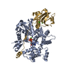
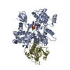
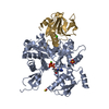
| Entry | Database: PDB / ID: 3a5n | ||||||
|---|---|---|---|---|---|---|---|
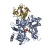
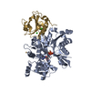
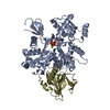
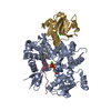
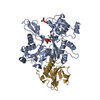
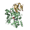
| Title | Crystal Structure of a Dictyostelium P109A Ca2+-Actin in Complex with Human Gelsolin Segment 1 | ||||||
 Components Components |
| ||||||
 Keywords Keywords | CONTRACTILE PROTEIN / ACTIN / ADP / HYDROLYSIS / ACTIN CAPPING / ACTIN-BINDING / CYTOSKELETON / ATP-BINDING / NUCLEOTIDE-BINDING / STRUCTURAL PROTEIN | ||||||
| Function / homology |  Function and homology information Function and homology informationintranuclear rod / leading edge of lamellipodium / phototaxis / phagolysosome / macropinocytic cup / striated muscle atrophy / regulation of establishment of T cell polarity / regulation of plasma membrane raft polarization / regulation of receptor clustering / positive regulation of keratinocyte apoptotic process ...intranuclear rod / leading edge of lamellipodium / phototaxis / phagolysosome / macropinocytic cup / striated muscle atrophy / regulation of establishment of T cell polarity / regulation of plasma membrane raft polarization / regulation of receptor clustering / positive regulation of keratinocyte apoptotic process / cell pole / renal protein absorption / positive regulation of protein processing in phagocytic vesicle / phosphatidylinositol 3-kinase catalytic subunit binding / positive regulation of actin nucleation / actin cap / myosin II binding / host-mediated suppression of symbiont invasion / cell projection assembly / hyperosmotic response / actin filament severing / early phagosome / barbed-end actin filament capping / actin polymerization or depolymerization / plasma membrane repair / actin filament depolymerization / actin filament capping / relaxation of cardiac muscle / Sensory processing of sound by outer hair cells of the cochlea / phagocytosis, engulfment / cardiac muscle cell contraction / hepatocyte apoptotic process / cortical actin cytoskeleton / myosin binding / cell leading edge / pseudopodium / mitotic cytokinesis / sarcoplasm / cilium assembly / Caspase-mediated cleavage of cytoskeletal proteins / endocytic vesicle / response to cAMP / phagocytosis / phagocytic cup / phagocytic vesicle / response to muscle stretch / vesicle-mediated transport / actin filament polymerization / phosphatidylinositol-4,5-bisphosphate binding / lipid droplet / actin filament organization / central nervous system development / actin filament / cellular response to type II interferon / protein destabilization / structural constituent of cytoskeleton / cell morphogenesis / Hydrolases; Acting on acid anhydrides; Acting on acid anhydrides to facilitate cellular and subcellular movement / chemotaxis / endocytosis / cell-cell junction / actin filament binding / lamellipodium / actin cytoskeleton / actin binding / secretory granule lumen / blood microparticle / cell cortex / amyloid fibril formation / ficolin-1-rich granule lumen / Amyloid fiber formation / focal adhesion / hydrolase activity / calcium ion binding / positive regulation of gene expression / Neutrophil degranulation / : / extracellular exosome / extracellular region / ATP binding / plasma membrane / cytosol / cytoplasm Similarity search - Function | ||||||
| Biological species |  HOMO SAPIENS (human) HOMO SAPIENS (human) | ||||||
| Method |  X-RAY DIFFRACTION / SYNCHROTRON / MOLECULAR REPLACEMENT / Resolution: 2.36 Å X-RAY DIFFRACTION / SYNCHROTRON / MOLECULAR REPLACEMENT / Resolution: 2.36 Å | ||||||
 Authors Authors | Murakami, K. / Yasunaga, T. / Noguchi, T.Q. / Uyeda, T.Q. / Wakabayashi, T. | ||||||
 Citation Citation | Journal: Cell / Year: 2010 Title: Structural basis for actin assembly, activation of ATP hydrolysis, and delayed phosphate release. Authors: Kenji Murakami / Takuo Yasunaga / Taro Q P Noguchi / Yuki Gomibuchi / Kien X Ngo / Taro Q P Uyeda / Takeyuki Wakabayashi /  Abstract: Assembled actin filaments support cellular signaling, intracellular trafficking, and cytokinesis. ATP hydrolysis triggered by actin assembly provides the structural cues for filament turnover in vivo. ...Assembled actin filaments support cellular signaling, intracellular trafficking, and cytokinesis. ATP hydrolysis triggered by actin assembly provides the structural cues for filament turnover in vivo. Here, we present the cryo-electron microscopic (cryo-EM) structure of filamentous actin (F-actin) in the presence of phosphate, with the visualization of some α-helical backbones and large side chains. A complete atomic model based on the EM map identified intermolecular interactions mediated by bound magnesium and phosphate ions. Comparison of the F-actin model with G-actin monomer crystal structures reveals a critical role for bending of the conserved proline-rich loop in triggering phosphate release following ATP hydrolysis. Crystal structures of G-actin show that mutations in this loop trap the catalytic site in two intermediate states of the ATPase cycle. The combined structural information allows us to propose a detailed molecular mechanism for the biochemical events, including actin polymerization and ATPase activation, critical for actin filament dynamics. | ||||||
| History |
|
- Structure visualization
| Structure viewer | Molecule: MolmilJmol/JSmol |
|---|
- Downloads & links
-Download
| PDBx/mmCIF format | 3a5n.cif.gz | 122.4 KB | Display | PDBx/mmCIF format |
|---|---|---|---|---|
| PDB format | pdb3a5n.ent.gz | 91.1 KB | Display | PDB format |
| PDBx/mmJSON format | 3a5n.json.gz | Tree view | PDBx/mmJSON format | |
| Others |  Other downloads Other downloads |
-Validation report
| Arichive directory | https://data.pdbj.org/pub/pdb/validation_reports/a5/3a5nftp://data.pdbj.org/pub/pdb/validation_reports/a5/3a5n | HTTPS FTP |
|---|
-Related structure data
| Related structure data |  1674C 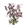 3a5lC 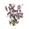 3a5mC 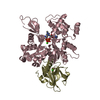 3a5oC  3g37C 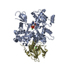 1c0gS C: citing same article ( S: Starting model for refinement |
|---|---|
| Similar structure data |


-Links
 PDBj
PDBj


- Assembly
| Deposited unit | 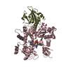
| ||||||||
|---|---|---|---|---|---|---|---|---|---|
| 1 |
| ||||||||
| Unit cell |
|
-Components
| #1: Protein | Mass: 14205.001 Da / Num. of mol.: 1 / Fragment: GELSOLIN-LIKE 1, residues 53-176 / Mutation: N35C Source method: isolated from a genetically manipulated source Source: (gene. exp.) HOMO SAPIENS (human) / Plasmid: PTIKL / Production host:  | ||||
|---|---|---|---|---|---|
| #2: Protein | Mass: 41432.203 Da / Num. of mol.: 1 / Mutation: P109A/E208A/R206A/E207A Source method: isolated from a genetically manipulated source Source: (gene. exp.) | ||||
| #3: Chemical |   Mass: 40.078 Da / Num. of mol.: 3 / Source method: obtained synthetically / Formula: Ca Mass: 40.078 Da / Num. of mol.: 3 / Source method: obtained synthetically / Formula: Ca#4: Chemical | ChemComp-ATP / |   Mass: 507.181 Da / Num. of mol.: 1 / Source method: obtained synthetically / Formula: C10H16N5O13P3 / Comment: ATP, energy-carrying molecule*YM Mass: 507.181 Da / Num. of mol.: 1 / Source method: obtained synthetically / Formula: C10H16N5O13P3 / Comment: ATP, energy-carrying molecule*YM#5: Water | ChemComp-HOH / |  Mass: 18.015 Da / Num. of mol.: 368 / Source method: isolated from a natural source / Formula: H2O Mass: 18.015 Da / Num. of mol.: 368 / Source method: isolated from a natural source / Formula: H2O |
-Experimental details
-Experiment
| Experiment | Method: X-RAY DIFFRACTION / Number of used crystals: 1 |
|---|
- Sample preparation
| Crystal | Density Matthews: 3.17 Å3/Da / Density % sol: 61.2 % |
|---|---|
| Crystal grow | Temperature: 293 K / Method: vapor diffusion, hanging drop / Details: VAPOR DIFFUSION, HANGING DROP, temperature 293K |
-Data collection
| Diffraction | Mean temperature: 100 K |
|---|---|
| Diffraction source | Source: SYNCHROTRON / Site: Photon Factory / Beamline: BL-5A / Wavelength: 1 Å |
| Detector | Type: ADSC QUANTUM 315 / Detector: CCD / Date: Jan 22, 2008 |
| Radiation | Protocol: SINGLE WAVELENGTH / Monochromatic (M) / Laue (L): M / Scattering type: x-ray |
| Radiation wavelength | Wavelength: 1 Å / Relative weight: 1 |
| Reflection | Resolution: 2.35→6 Å / Num. obs: 29796 / Biso Wilson estimate: 34.3 Å2 / Rmerge(I) obs: 0.183 |
| Reflection shell | Resolution: 2.35→2.44 Å / Rmerge(I) obs: 0.292 / Mean I/σ(I) obs: 3.97 |
- Processing
| Software |
| ||||||||||||||||||||||||||||||||||||||||||||||||||||||||||||||||||||||||||||||||||||||||||||||||||||||||||||||||||||||||||||||||||||||||||||||||||||||||||||||||||||||||||
|---|---|---|---|---|---|---|---|---|---|---|---|---|---|---|---|---|---|---|---|---|---|---|---|---|---|---|---|---|---|---|---|---|---|---|---|---|---|---|---|---|---|---|---|---|---|---|---|---|---|---|---|---|---|---|---|---|---|---|---|---|---|---|---|---|---|---|---|---|---|---|---|---|---|---|---|---|---|---|---|---|---|---|---|---|---|---|---|---|---|---|---|---|---|---|---|---|---|---|---|---|---|---|---|---|---|---|---|---|---|---|---|---|---|---|---|---|---|---|---|---|---|---|---|---|---|---|---|---|---|---|---|---|---|---|---|---|---|---|---|---|---|---|---|---|---|---|---|---|---|---|---|---|---|---|---|---|---|---|---|---|---|---|---|---|---|---|---|---|---|---|---|
| Refinement | Method to determine structure: MOLECULAR REPLACEMENT Starting model: PDB ENTRY 1C0G Resolution: 2.36→6 Å / Cor.coef. Fo:Fc: 0.936 / Cor.coef. Fo:Fc free: 0.926 / Occupancy max: 1 / Occupancy min: 0 / SU B: 5.878 / SU ML: 0.14 / Cross valid method: THROUGHOUT / ESU R: 0.349 / ESU R Free: 0.211 / Stereochemistry target values: MAXIMUM LIKELIHOOD / Details: HYDROGENS HAVE BEEN ADDED IN THE RIDING POSITIONS
| ||||||||||||||||||||||||||||||||||||||||||||||||||||||||||||||||||||||||||||||||||||||||||||||||||||||||||||||||||||||||||||||||||||||||||||||||||||||||||||||||||||||||||
| Solvent computation | Ion probe radii: 0.8 Å / Shrinkage radii: 0.8 Å / VDW probe radii: 1.2 Å / Solvent model: MASK | ||||||||||||||||||||||||||||||||||||||||||||||||||||||||||||||||||||||||||||||||||||||||||||||||||||||||||||||||||||||||||||||||||||||||||||||||||||||||||||||||||||||||||
| Displacement parameters | Biso max: 87.86 Å2 / Biso mean: 26.887 Å2 / Biso min: 6.63 Å2
| ||||||||||||||||||||||||||||||||||||||||||||||||||||||||||||||||||||||||||||||||||||||||||||||||||||||||||||||||||||||||||||||||||||||||||||||||||||||||||||||||||||||||||
| Refinement step | Cycle: LAST / Resolution: 2.36→6 Å
| ||||||||||||||||||||||||||||||||||||||||||||||||||||||||||||||||||||||||||||||||||||||||||||||||||||||||||||||||||||||||||||||||||||||||||||||||||||||||||||||||||||||||||
| Refine LS restraints |
| ||||||||||||||||||||||||||||||||||||||||||||||||||||||||||||||||||||||||||||||||||||||||||||||||||||||||||||||||||||||||||||||||||||||||||||||||||||||||||||||||||||||||||
| LS refinement shell | Resolution: 2.36→2.42 Å / Total num. of bins used: 20
|
