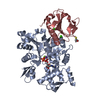
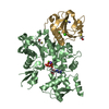
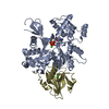
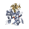
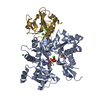
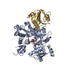
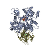
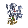
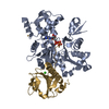
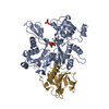
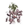
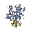
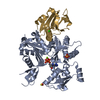
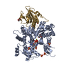
[English] 日本語
Yorodumi
- PDB-5mvv: Crystal structure of Plasmodium falciparum actin I- gelsolin segm... -

+
Open data


ID or keywords:

Loading...

-
Basic information

Entry
Database: PDB / ID: 5mvv
TitleCrystal structure of Plasmodium falciparum actin I- gelsolin segment 1 -CdATP complex
Components
  • Actin-1
  • Gelsolin
KeywordsSTRUCTURAL PROTEIN / experimental phasing / cadmium / atp / actin / SAD phasing
Function / homology
Function and homology information


glial filament / plastid inheritance / schizogony / apical ectoplasmic specialization / basal ectoplasmic specialization / Caspase-mediated cleavage of cytoskeletal proteins / symbiont-mediated actin polymerization-dependent cell-to-cell migration in host / HSP90 chaperone cycle for steroid hormone receptors (SHR) in the presence of ligand / striated muscle atrophy / regulation of establishment of T cell polarity ...glial filament / plastid inheritance / schizogony / apical ectoplasmic specialization / basal ectoplasmic specialization / Caspase-mediated cleavage of cytoskeletal proteins / symbiont-mediated actin polymerization-dependent cell-to-cell migration in host / HSP90 chaperone cycle for steroid hormone receptors (SHR) in the presence of ligand / striated muscle atrophy / regulation of establishment of T cell polarity / regulation of plasma membrane raft polarization / regulation of receptor clustering / renal protein absorption / positive regulation of keratinocyte apoptotic process / positive regulation of protein processing in phagocytic vesicle / positive regulation of actin nucleation / phosphatidylinositol 3-kinase catalytic subunit binding / actin cap / Neutrophil degranulation / regulation of podosome assembly / sequestering of actin monomers / myosin II binding / negative regulation of viral entry into host cell / entry into host cell by a symbiont-containing vacuole / actin filament severing / actin filament capping / actin filament depolymerization / actin polymerization or depolymerization / barbed-end actin filament capping / cell projection assembly / cardiac muscle cell contraction / podosome / relaxation of cardiac muscle / positive regulation of p38MAPK cascade / phagocytosis, engulfment / positive regulation of cardiac muscle hypertrophy / hepatocyte apoptotic process / sarcoplasm / cilium assembly / vesicle-mediated transport / phagocytic vesicle / cytoskeleton organization / ruffle / phosphatidylinositol-4,5-bisphosphate binding / response to muscle stretch / cellular response to cadmium ion / actin filament polymerization / Neutrophil degranulation / central nervous system development / actin filament / Hydrolases; Acting on acid anhydrides; Acting on acid anhydrides to facilitate cellular and subcellular movement / protein destabilization / structural constituent of cytoskeleton / cellular response to type II interferon / actin filament binding / actin cytoskeleton / lamellipodium / myelin sheath / actin binding / actin cytoskeleton organization / amyloid fibril formation / calcium ion binding / perinuclear region of cytoplasm / ATP hydrolysis activity / protein-containing complex / extracellular space / extracellular region / ATP binding / nucleus / plasma membrane / cytosol / cytoplasm
Similarity search - Function
Villin/Gelsolin / Gelsolin homology domain / Severin / Severin / Gelsolin-like domain / Gelsolin repeat / ADF-H/Gelsolin-like domain superfamily / ATPase, substrate binding domain, subdomain 4 / Actin; Chain A, domain 4 / ATPase, nucleotide binding domain ...Villin/Gelsolin / Gelsolin homology domain / Severin / Severin / Gelsolin-like domain / Gelsolin repeat / ADF-H/Gelsolin-like domain superfamily / ATPase, substrate binding domain, subdomain 4 / Actin; Chain A, domain 4 / ATPase, nucleotide binding domain / Actins signature 1. / Actin, conserved site / Actins signature 2. / Actin/actin-like conserved site / Actins and actin-related proteins signature. / Actin / Actin family / Actin / ATPase, nucleotide binding domain / Nucleotidyltransferase; domain 5 / Alpha-Beta Complex / 2-Layer Sandwich / 3-Layer(aba) Sandwich / Alpha Beta
Similarity search - Domain/homology
ADENOSINE-5'-TRIPHOSPHATE / : / THIOCYANATE ION / Gelsolin / Actin-1
Similarity search - Component
Biological speciesPlasmodium falciparum (malaria parasite P. falciparum)
Mus musculus (house mouse)
MethodX-RAY DIFFRACTION / SYNCHROTRON / SAD / Resolution: 1.4 Å
AuthorsPanneerselvam, S. / Kumpula, E.-P. / Kursula, I. / Burkhardt, A. / Meents, A.
CitationJournal: Acta Crystallogr D Struct Biol / Year: 2017
Title: Rapid cadmium SAD phasing at the standard wavelength (1 angstrom ).
Authors: Panneerselvam, S. / Kumpula, E.P. / Kursula, I. / Burkhardt, A. / Meents, A.
History
DepositionJan 17, 2017Deposition site: PDBE / Processing site: PDBE
Revision 1.0Jul 12, 2017Provider: repository / Type: Initial release
Revision 2.0Jul 19, 2017Group: Atomic model / Category: atom_site / Item: _atom_site.occupancy
Revision 2.1Aug 2, 2017Group: Database references / Category: citation / citation_author
Item: _citation.country / _citation.journal_abbrev ..._citation.country / _citation.journal_abbrev / _citation.journal_id_ASTM / _citation.journal_id_CSD / _citation.journal_id_ISSN / _citation.pdbx_database_id_PubMed / _citation.title / _citation_author.name
Revision 2.2Oct 16, 2019Group: Data collection / Category: reflns_shell
Revision 2.3May 8, 2024Group: Data collection / Database references / Derived calculations
Category: chem_comp_atom / chem_comp_bond ...chem_comp_atom / chem_comp_bond / database_2 / pdbx_struct_conn_angle / struct_conn
Item: _database_2.pdbx_DOI / _database_2.pdbx_database_accession ..._database_2.pdbx_DOI / _database_2.pdbx_database_accession / _pdbx_struct_conn_angle.ptnr1_auth_asym_id / _pdbx_struct_conn_angle.ptnr1_auth_comp_id / _pdbx_struct_conn_angle.ptnr1_auth_seq_id / _pdbx_struct_conn_angle.ptnr1_label_alt_id / _pdbx_struct_conn_angle.ptnr1_label_asym_id / _pdbx_struct_conn_angle.ptnr1_label_atom_id / _pdbx_struct_conn_angle.ptnr1_label_comp_id / _pdbx_struct_conn_angle.ptnr1_label_seq_id / _pdbx_struct_conn_angle.ptnr2_auth_asym_id / _pdbx_struct_conn_angle.ptnr2_auth_comp_id / _pdbx_struct_conn_angle.ptnr2_auth_seq_id / _pdbx_struct_conn_angle.ptnr2_label_asym_id / _pdbx_struct_conn_angle.ptnr2_label_atom_id / _pdbx_struct_conn_angle.ptnr2_label_comp_id / _pdbx_struct_conn_angle.ptnr3_auth_asym_id / _pdbx_struct_conn_angle.ptnr3_auth_comp_id / _pdbx_struct_conn_angle.ptnr3_auth_seq_id / _pdbx_struct_conn_angle.ptnr3_label_alt_id / _pdbx_struct_conn_angle.ptnr3_label_asym_id / _pdbx_struct_conn_angle.ptnr3_label_atom_id / _pdbx_struct_conn_angle.ptnr3_label_comp_id / _pdbx_struct_conn_angle.ptnr3_label_seq_id / _pdbx_struct_conn_angle.value / _struct_conn.pdbx_dist_value / _struct_conn.pdbx_ptnr1_label_alt_id / _struct_conn.ptnr1_auth_asym_id / _struct_conn.ptnr1_auth_comp_id / _struct_conn.ptnr1_auth_seq_id / _struct_conn.ptnr1_label_asym_id / _struct_conn.ptnr1_label_atom_id / _struct_conn.ptnr1_label_comp_id / _struct_conn.ptnr1_label_seq_id / _struct_conn.ptnr2_auth_asym_id / _struct_conn.ptnr2_auth_comp_id / _struct_conn.ptnr2_auth_seq_id / _struct_conn.ptnr2_label_asym_id / _struct_conn.ptnr2_label_atom_id / _struct_conn.ptnr2_label_comp_id / _struct_conn.ptnr2_label_seq_id

-
Structure visualization

Structure viewerMolecule:
MolmilJmol/JSmol

Downloads & links

-
Assembly

Deposited unit
A: Actin-1
G: Gelsolin
hetero molecules


Theoretical massNumber of molelcules
Total (without water)57,2429
Polymers56,2602
Non-polymers9837
Water8,143452
1


  • Idetical with deposited unit
  • defined by author&software
TypeNameSymmetry operationNumber
identity operation1_555x,y,z1
Buried area3950 Å2
ΔGint-66 kcal/mol
Surface area20380 Å2
MethodPISA
Unit cell
Length a, b, c (Å)110.420, 68.920, 71.550
Angle α, β, γ (deg.)90.00, 90.00, 90.00
Int Tables number18
Space group name H-MP21212

-
Components

-
Protein , 2 types, 2 molecules AG

#1: Protein Actin-1 / Actin I


Mass: 42047.676 Da / Num. of mol.: 1
Source method: isolated from a genetically manipulated source
Source: (gene. exp.) Plasmodium falciparum (isolate 3D7) (eukaryote)
Strain: isolate 3D7 / Gene: PFL2215w / Production host: 
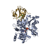
Spodoptera frugiperda (fall armyworm) / Strain (production host): Sf21 / References: UniProt: Q8I4X0
#2: Protein Gelsolin / Actin-depolymerizing factor / ADF / Brevin


Mass: 14211.929 Da / Num. of mol.: 1
Source method: isolated from a genetically manipulated source
Source: (gene. exp.) Mus musculus (house mouse) / Gene: Gsn, Gsb / Plasmid: pETM22 / Production host: Escherichia coli (E. coli) / Strain (production host): BL21(DE3) / References: UniProt: P13020

-
Non-polymers , 5 types, 459 molecules

#3: Chemical ChemComp-ATP / ADENOSINE-5'-TRIPHOSPHATE


Mass: 507.181 Da / Num. of mol.: 1 / Source method: obtained synthetically / Formula: C10H16N5O13P3 / Comment: ATP, energy-carrying molecule*YM
#4: Chemical ChemComp-SCN / THIOCYANATE ION


Mass: 58.082 Da / Num. of mol.: 1 / Source method: obtained synthetically / Formula: CNS
#5: Chemical ChemComp-CD / CADMIUM ION


Mass: 112.411 Da / Num. of mol.: 3 / Source method: obtained synthetically / Formula: Cd
#6: Chemical ChemComp-CA / CALCIUM ION


Mass: 40.078 Da / Num. of mol.: 2 / Source method: obtained synthetically / Formula: Ca
#7: Water ChemComp-HOH / water


Mass: 18.015 Da / Num. of mol.: 452 / Source method: isolated from a natural source / Formula: H2O

-
Experimental details

-
Experiment

ExperimentMethod: X-RAY DIFFRACTION / Number of used crystals: 1

-
Sample preparation

CrystalDensity Matthews: 2.42 Å3/Da / Density % sol: 49.17 %
Crystal growTemperature: 293.15 K / Method: vapor diffusion, sitting drop / pH: 5.9
Details: 14 or 15% PEG3350, 0.2M potassium thiocyanate, 0.1M bis-tris pH 5.9. Drops were streak-seeded from crystals with calcium.

-
Data collection

DiffractionMean temperature: 80 K
Diffraction sourceSource: SYNCHROTRON / Site: PETRA III, DESY / Beamline: P11 / Wavelength: 0.9806 Å
DetectorType: DECTRIS PILATUS 6M-F / Detector: PIXEL / Date: Jun 19, 2015
RadiationProtocol: SINGLE WAVELENGTH / Monochromatic (M) / Laue (L): M / Scattering type: x-ray
Radiation wavelengthWavelength: 0.9806 Å / Relative weight: 1
ReflectionResolution: 1.4→45 Å / Num. obs: 72676 / % possible obs: 100 % / Redundancy: 12.6 % / Net I/σ(I): 33.81

-
Processing

Software
NameVersionClassification
REFMAC5.8.0158refinement
XDSdata reduction
XSCALEdata scaling
SHELXDEphasing
RefinementMethod to determine structure: SAD / Resolution: 1.4→71.55 Å / Cor.coef. Fo:Fc: 0.977 / Cor.coef. Fo:Fc free: 0.963 / SU B: 1.677 / SU ML: 0.03 / Cross valid method: THROUGHOUT / ESU R: 0.05 / ESU R Free: 0.048 / Stereochemistry target values: MAXIMUM LIKELIHOOD / Details: HYDROGENS HAVE BEEN ADDED IN THE RIDING POSITIONS
RfactorNum. reflection% reflectionSelection details
Rfree0.15853 2177 2 %RANDOM
Rwork0.12869 ---
obs0.12948 105710 99.95 %-
Solvent computationIon probe radii: 0.9 Å / Shrinkage radii: 0.9 Å / VDW probe radii: 1.2 Å / Solvent model: MASK
Displacement parametersBiso mean: 18.695 Å2
Baniso -1Baniso -2Baniso -3
1--0.39 Å2-0 Å20 Å2
2--0.36 Å2-0 Å2
3---0.03 Å2
Refinement stepCycle: 1 / Resolution: 1.4→71.55 Å
ProteinNucleic acidLigandSolventTotal
Num. atoms3738 0 39 452 4229
Refine LS restraints
Refine-IDTypeDev idealDev ideal targetNumber
X-RAY DIFFRACTIONr_bond_refined_d0.0160.0194447
X-RAY DIFFRACTIONr_bond_other_d0.0020.024013
X-RAY DIFFRACTIONr_angle_refined_deg1.7841.9776107
X-RAY DIFFRACTIONr_angle_other_deg1.0339388
X-RAY DIFFRACTIONr_dihedral_angle_1_deg5.9915584
X-RAY DIFFRACTIONr_dihedral_angle_2_deg34.85824.3200
X-RAY DIFFRACTIONr_dihedral_angle_3_deg12.1715762
X-RAY DIFFRACTIONr_dihedral_angle_4_deg14.9431525
X-RAY DIFFRACTIONr_chiral_restr0.1070.2646
X-RAY DIFFRACTIONr_gen_planes_refined0.010.0215191
X-RAY DIFFRACTIONr_gen_planes_other0.0040.02940
X-RAY DIFFRACTIONr_nbd_refined
X-RAY DIFFRACTIONr_nbd_other
X-RAY DIFFRACTIONr_nbtor_refined
X-RAY DIFFRACTIONr_nbtor_other
X-RAY DIFFRACTIONr_xyhbond_nbd_refined
X-RAY DIFFRACTIONr_xyhbond_nbd_other
X-RAY DIFFRACTIONr_metal_ion_refined
X-RAY DIFFRACTIONr_metal_ion_other
X-RAY DIFFRACTIONr_symmetry_vdw_refined
X-RAY DIFFRACTIONr_symmetry_vdw_other
X-RAY DIFFRACTIONr_symmetry_hbond_refined
X-RAY DIFFRACTIONr_symmetry_hbond_other
X-RAY DIFFRACTIONr_symmetry_metal_ion_refined
X-RAY DIFFRACTIONr_symmetry_metal_ion_other
X-RAY DIFFRACTIONr_mcbond_it2.1491.6132193
X-RAY DIFFRACTIONr_mcbond_other2.1421.6132191
X-RAY DIFFRACTIONr_mcangle_it2.8392.4142824
X-RAY DIFFRACTIONr_mcangle_other2.8382.4142825
X-RAY DIFFRACTIONr_scbond_it2.3241.9242254
X-RAY DIFFRACTIONr_scbond_other2.3211.9242253
X-RAY DIFFRACTIONr_scangle_it
X-RAY DIFFRACTIONr_scangle_other2.9212.763282
X-RAY DIFFRACTIONr_long_range_B_refined4.05420.1225153
X-RAY DIFFRACTIONr_long_range_B_other3.98419.9945123
X-RAY DIFFRACTIONr_rigid_bond_restr2.52238460
X-RAY DIFFRACTIONr_sphericity_free30.7495321
X-RAY DIFFRACTIONr_sphericity_bonded12.17858473
LS refinement shellResolution: 1.4→1.436 Å / Total num. of bins used: 20
RfactorNum. reflection% reflection
Rwork0.154 7853 -
Rfree-0 -
obs--99.83 %

+
About Yorodumi

-
News

-
Feb 9, 2022. New format data for meta-information of EMDB entries

New format data for meta-information of EMDB entries

  • Version 3 of the EMDB header file is now the official format.
  • The previous official version 1.9 will be removed from the archive.

Related info.:EMDB header

External links:wwPDB to switch to version 3 of the EMDB data model

-
Aug 12, 2020. Covid-19 info

Covid-19 info

URL: https://pdbjlc1.pdbj.org/emnavi/covid19.php

New page: Covid-19 featured information page in EM Navigator.

Related info.:Covid-19 info / Mar 5, 2020. Novel coronavirus structure data

+
Mar 5, 2020. Novel coronavirus structure data

Novel coronavirus structure data

Related info.:Yorodumi Speices / Aug 12, 2020. Covid-19 info

External links:COVID-19 featured content - PDBj / Molecule of the Month (242):Coronavirus Proteases

+
Jan 31, 2019. EMDB accession codes are about to change! (news from PDBe EMDB page)

EMDB accession codes are about to change! (news from PDBe EMDB page)

  • The allocation of 4 digits for EMDB accession codes will soon come to an end. Whilst these codes will remain in use, new EMDB accession codes will include an additional digit and will expand incrementally as the available range of codes is exhausted. The current 4-digit format prefixed with “EMD-” (i.e. EMD-XXXX) will advance to a 5-digit format (i.e. EMD-XXXXX), and so on. It is currently estimated that the 4-digit codes will be depleted around Spring 2019, at which point the 5-digit format will come into force.
  • The EM Navigator/Yorodumi systems omit the EMD- prefix.

Related info.:Q: What is EMD? / ID/Accession-code notation in Yorodumi/EM Navigator

External links:EMDB Accession Codes are Changing Soon! / Contact to PDBj

+
Jul 12, 2017. Major update of PDB

Major update of PDB

  • wwPDB released updated PDB data conforming to the new PDBx/mmCIF dictionary.
  • This is a major update changing the version number from 4 to 5, and with Remediation, in which all the entries are updated.
  • In this update, many items about electron microscopy experimental information are reorganized (e.g. em_software).
  • Now, EM Navigator and Yorodumi are based on the updated data.

External links:wwPDB Remediation / Enriched Model Files Conforming to OneDep Data Standards Now Available in the PDB FTP Archive

-
Yorodumi

Thousand views of thousand structures

  • Yorodumi is a browser for structure data from EMDB, PDB, SASBDB, etc.
  • This page is also the successor to EM Navigator detail page, and also detail information page/front-end page for Omokage search.
  • The word "yorodu" (or yorozu) is an old Japanese word meaning "ten thousand". "mi" (miru) is to see.

Related info.:EMDB / PDB / SASBDB / Comparison of 3 databanks / Yorodumi Search / Aug 31, 2016. New EM Navigator & Yorodumi / Yorodumi Papers / Jmol/JSmol / Function and homology information / Changes in new EM Navigator and Yorodumi

Read more