 Movie
Movie Controller

[English] 日本語
 Yorodumi
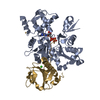
- PDB-1nlv: Crystal Structure Of Dictyostelium Discoideum Actin Complexed Wit... -
+ Open data

- Basic information
| Entry | Database: PDB / ID: 1nlv | ||||||
|---|---|---|---|---|---|---|---|
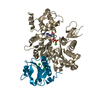
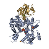
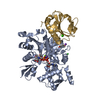
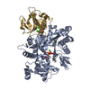
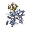
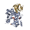
| Title | Crystal Structure Of Dictyostelium Discoideum Actin Complexed With Ca ATP And Human Gelsolin Segment 1 | ||||||
 Components Components |
| ||||||
 Keywords Keywords | STRUCTURAL PROTEIN / actin / gelsolin / cytoskeleton / cell motility | ||||||
| Function / homology |  Function and homology information Function and homology informationintranuclear rod / leading edge of lamellipodium / phototaxis / phagolysosome / macropinocytic cup / striated muscle atrophy / regulation of establishment of T cell polarity / regulation of plasma membrane raft polarization / regulation of receptor clustering / positive regulation of keratinocyte apoptotic process ...intranuclear rod / leading edge of lamellipodium / phototaxis / phagolysosome / macropinocytic cup / striated muscle atrophy / regulation of establishment of T cell polarity / regulation of plasma membrane raft polarization / regulation of receptor clustering / positive regulation of keratinocyte apoptotic process / renal protein absorption / positive regulation of protein processing in phagocytic vesicle / phosphatidylinositol 3-kinase catalytic subunit binding / cell pole / positive regulation of actin nucleation / actin cap / myosin II binding / host-mediated suppression of symbiont invasion / actin filament severing / barbed-end actin filament capping / early phagosome / cell projection assembly / plasma membrane repair / actin polymerization or depolymerization / actin filament depolymerization / actin filament capping / hyperosmotic response / relaxation of cardiac muscle / Sensory processing of sound by outer hair cells of the cochlea / phagocytosis, engulfment / cardiac muscle cell contraction / cortical actin cytoskeleton / myosin binding / hepatocyte apoptotic process / cell leading edge / pseudopodium / mitotic cytokinesis / sarcoplasm / cilium assembly / Caspase-mediated cleavage of cytoskeletal proteins / endocytic vesicle / response to cAMP / phagocytosis / phagocytic cup / phagocytic vesicle / response to muscle stretch / vesicle-mediated transport / actin filament polymerization / phosphatidylinositol-4,5-bisphosphate binding / lipid droplet / actin filament organization / central nervous system development / actin filament / structural constituent of cytoskeleton / cellular response to type II interferon / protein destabilization / Hydrolases; Acting on acid anhydrides; Acting on acid anhydrides to facilitate cellular and subcellular movement / cell morphogenesis / chemotaxis / endocytosis / cell-cell junction / actin filament binding / lamellipodium / actin cytoskeleton / actin binding / secretory granule lumen / blood microparticle / cell cortex / amyloid fibril formation / ficolin-1-rich granule lumen / Amyloid fiber formation / focal adhesion / hydrolase activity / calcium ion binding / Neutrophil degranulation / positive regulation of gene expression / : / extracellular exosome / extracellular region / ATP binding / plasma membrane / cytoplasm / cytosol Similarity search - Function | ||||||
| Biological species |  Homo sapiens (human) Homo sapiens (human) | ||||||
| Method |  X-RAY DIFFRACTION / SYNCHROTRON / MOLECULAR REPLACEMENT / Resolution: 1.8 Å X-RAY DIFFRACTION / SYNCHROTRON / MOLECULAR REPLACEMENT / Resolution: 1.8 Å | ||||||
 Authors Authors | Vorobiev, S.M. / Welti, S. / Condeelis, J. / Almo, S.C. | ||||||
 Citation Citation | Journal: Proc.Natl.Acad.Sci.USA / Year: 2003 Title: The Structure of Non-Vertebrate Actin: Implications For The ATP Hydrolytic Mechanism Authors: Vorobiev, S.M. / Strokopytov, B. / Drubin, D.G. / Frieden, C. / Ono, S. / Condeelis, J. / Rubenstein, P.A. / Almo, S.C. | ||||||
| History |
|
- Structure visualization
| Structure viewer | Molecule: MolmilJmol/JSmol |
|---|
- Downloads & links
-Download
| PDBx/mmCIF format | 1nlv.cif.gz | 118.9 KB | Display | PDBx/mmCIF format |
|---|---|---|---|---|
| PDB format | pdb1nlv.ent.gz | 89 KB | Display | PDB format |
| PDBx/mmJSON format | 1nlv.json.gz | Tree view | PDBx/mmJSON format | |
| Others |  Other downloads Other downloads |
-Validation report
| Arichive directory | https://data.pdbj.org/pub/pdb/validation_reports/nl/1nlvftp://data.pdbj.org/pub/pdb/validation_reports/nl/1nlv | HTTPS FTP |
|---|
-Related structure data
| Related structure data | 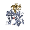 1d4xC  1nm1C  1nmdC 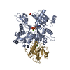 1yagC 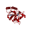 1dagS C: citing same article ( S: Starting model for refinement |
|---|---|
| Similar structure data |


-Links
 PDBj
PDBj


- Assembly
| Deposited unit | 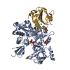
| |||||||||
|---|---|---|---|---|---|---|---|---|---|---|
| 1 |
| |||||||||
| 2 | 
| |||||||||
| Unit cell |
| |||||||||
| Components on special symmetry positions |
|
-Components
-Protein , 2 types, 2 molecules AG
| #1: Protein | Mass: 41647.410 Da / Num. of mol.: 1 / Source method: isolated from a natural source / Source: (natural) |
|---|---|
| #2: Protein | Mass: 14071.831 Da / Num. of mol.: 1 / Fragment: DOMAIN 1 Source method: isolated from a genetically manipulated source Source: (gene. exp.) Homo sapiens (human) / Gene: GSN / Plasmid: pMW172 / Species (production host): Escherichia coli / Production host:  |
-Non-polymers , 5 types, 337 molecules 


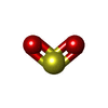

| #3: Chemical |  Mass: 40.078 Da / Num. of mol.: 2 / Source method: obtained synthetically / Formula: Ca Mass: 40.078 Da / Num. of mol.: 2 / Source method: obtained synthetically / Formula: Ca#4: Chemical |  Mass: 96.063 Da / Num. of mol.: 2 / Source method: obtained synthetically / Formula: SO4 Mass: 96.063 Da / Num. of mol.: 2 / Source method: obtained synthetically / Formula: SO4#5: Chemical | ChemComp-ATP / |  Mass: 507.181 Da / Num. of mol.: 1 / Source method: obtained synthetically / Formula: C10H16N5O13P3 / Comment: ATP, energy-carrying molecule*YM Mass: 507.181 Da / Num. of mol.: 1 / Source method: obtained synthetically / Formula: C10H16N5O13P3 / Comment: ATP, energy-carrying molecule*YM#6: Chemical | ChemComp-SO2 / |  Mass: 64.064 Da / Num. of mol.: 1 / Source method: obtained synthetically / Formula: O2S Mass: 64.064 Da / Num. of mol.: 1 / Source method: obtained synthetically / Formula: O2S#7: Water | ChemComp-HOH / | Mass: 18.015 Da / Num. of mol.: 331 / Source method: isolated from a natural source / Formula: H2O |
|---|
-Experimental details
-Experiment
| Experiment | Method: X-RAY DIFFRACTION / Number of used crystals: 1 |
|---|
- Sample preparation
| Crystal | Density Matthews: 3.03 Å3/Da / Density % sol: 59.38 % |
|---|---|
| Crystal grow | Temperature: 291 K / Method: vapor diffusion, hanging drop / pH: 7.5 Details: lithium chloride, magnesium chloride, HEPES, ATP, pH 7.5, VAPOR DIFFUSION, HANGING DROP, temperature 291K |
-Data collection
| Diffraction | Mean temperature: 95 K |
|---|---|
| Diffraction source | Source: SYNCHROTRON / Site: NSLS  / Beamline: X9B / Wavelength: 1.07018 Å / Beamline: X9B / Wavelength: 1.07018 Å |
| Detector | Type: ADSC QUANTUM 4 / Detector: CCD / Date: Apr 28, 1999 |
| Radiation | Protocol: SINGLE WAVELENGTH / Monochromatic (M) / Laue (L): M / Scattering type: x-ray |
| Radiation wavelength | Wavelength: 1.07018 Å / Relative weight: 1 |
| Reflection | Resolution: 1.8→23 Å / Num. all: 61207 / Num. obs: 61207 / % possible obs: 99.9 % / Observed criterion σ(I): 0 / Redundancy: 4.14 % |
| Reflection shell | Resolution: 1.8→1.86 Å / Rmerge(I) obs: 0.338 / Num. unique all: 6067 / % possible all: 99.4 |
- Processing
| Software |
| ||||||||||||||||
|---|---|---|---|---|---|---|---|---|---|---|---|---|---|---|---|---|---|
| Refinement | Method to determine structure: MOLECULAR REPLACEMENT Starting model: PDB ENTRY 1DAG Resolution: 1.8→23 Å / Cross valid method: THROUGHOUT / σ(F): 0 / Stereochemistry target values: Engh & Huber
| ||||||||||||||||
| Refinement step | Cycle: LAST / Resolution: 1.8→23 Å
|
