 Movie
Movie Controller

[English] 日本語
 Yorodumi
- PDB-1zee: X-Ray Crystal Structure of Protein SO4414 from Shewanella oneiden... -
+ Open data

- Basic information
| Entry | Database: PDB / ID: 1zee | ||||||
|---|---|---|---|---|---|---|---|
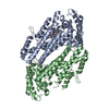
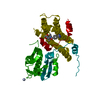
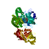
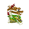
| Title | X-Ray Crystal Structure of Protein SO4414 from Shewanella oneidensis. Northeast Structural Genomics Consortium Target SoR52. | ||||||
 Components Components | hypothetical protein SO4414 | ||||||
 Keywords Keywords | UNKNOWN FUNCTION / all alpha-protein. / Structural Genomics / PSI / Protein Structure Initiative / Northeast Structural Genomics Consortium / NESG | ||||||
| Function / homology |  Function and homology information Function and homology informationtryptophan 2,3-dioxygenase / L-tryptophan 2,3-dioxygenase activity / : / heme binding / metal ion binding Similarity search - Function | ||||||
| Biological species |  Shewanella oneidensis (bacteria) Shewanella oneidensis (bacteria) | ||||||
| Method |  X-RAY DIFFRACTION / SYNCHROTRON / SAD / Resolution: 2.31 Å X-RAY DIFFRACTION / SYNCHROTRON / SAD / Resolution: 2.31 Å | ||||||
 Authors Authors | Forouhar, F. / Abashidze, M. / Vorobiev, S.M. / Conover, K. / Acton, T.B. / Montelione, G.T. / Hunt, J.F. / Tong, L. / Northeast Structural Genomics Consortium (NESG) | ||||||
 Citation Citation | Journal: To be Published Title: Crystal Structure of the Hypothetical Protein SO4414 from Shewanella oneidensis, NESG Target SoR52 Authors: Forouhar, F. / Abashidze, M. / Vorobiev, S.M. / Conover, K. / Acton, T.B. / Montelione, G.T. / Hunt, J.F. / Tong, L. | ||||||
| History |
|
- Structure visualization
| Structure viewer | Molecule: MolmilJmol/JSmol |
|---|
- Downloads & links
-Download
| PDBx/mmCIF format | 1zee.cif.gz | 161.6 KB | Display | PDBx/mmCIF format |
|---|---|---|---|---|
| PDB format | pdb1zee.ent.gz | 129.2 KB | Display | PDB format |
| PDBx/mmJSON format | 1zee.json.gz | Tree view | PDBx/mmJSON format | |
| Others |  Other downloads Other downloads |
-Validation report
| Arichive directory | https://data.pdbj.org/pub/pdb/validation_reports/ze/1zeeftp://data.pdbj.org/pub/pdb/validation_reports/ze/1zee | HTTPS FTP |
|---|
-Related structure data
| Similar structure data | |
|---|---|
| Other databases |


-Links
 PDBj
PDBj- Assembly
| Deposited unit | 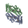
| ||||||||
|---|---|---|---|---|---|---|---|---|---|
| 1 |
| ||||||||
| 2 | 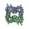
| ||||||||
| Unit cell |
|
-Components
| #1: Protein | Mass: 46745.523 Da / Num. of mol.: 2 Source method: isolated from a genetically manipulated source Source: (gene. exp.) Shewanella oneidensis (bacteria) / Strain: MR-1 / Gene: GeneID:1172013 / Plasmid: pET21 / Species (production host): Escherichia coli / Production host: Keywords: Substitution of Met residues by Seleno-Met residues and addition of C-tag (LEHHHHHH). SO4414References: UniProt: Q8E972 #2: Water | ChemComp-HOH / |  Mass: 18.015 Da / Num. of mol.: 309 / Source method: isolated from a natural source / Formula: H2O Mass: 18.015 Da / Num. of mol.: 309 / Source method: isolated from a natural source / Formula: H2OHas protein modification | Y | |
|---|
-Experimental details
-Experiment
| Experiment | Method: X-RAY DIFFRACTION / Number of used crystals: 1 |
|---|
- Sample preparation
| Crystal | Density Matthews: 2.36 Å3/Da / Density % sol: 47 % |
|---|---|
| Crystal grow | Temperature: 293 K / Method: vapor diffusion, hanging drop / pH: 7.5 Details: 18% PEG3350, 150 mM lithium sulfate, and 5 mM DTT., pH 7.5, VAPOR DIFFUSION, HANGING DROP, temperature 293K |
-Data collection
| Diffraction | Mean temperature: 100 K |
|---|---|
| Diffraction source | Source: SYNCHROTRON / Site: NSLS  / Beamline: X4A / Wavelength: 0.97915 / Wavelength: 0.97915 Å / Beamline: X4A / Wavelength: 0.97915 / Wavelength: 0.97915 Å |
| Detector | Type: ADSC QUANTUM 4 / Detector: CCD / Date: Mar 16, 2005 / Details: mirrors |
| Radiation | Monochromator: Si 111 CHANNEL / Protocol: SINGLE WAVELENGTH / Monochromatic (M) / Laue (L): M / Scattering type: x-ray |
| Radiation wavelength | Wavelength: 0.97915 Å / Relative weight: 1 |
| Reflection | Resolution: 2.3→30 Å / Num. all: 72798 / Num. obs: 70105 / % possible obs: 96.5 % / Observed criterion σ(F): 0 / Observed criterion σ(I): 0 / Redundancy: 4.9 % / Biso Wilson estimate: 28.9 Å2 / Rmerge(I) obs: 0.066 / Rsym value: 0.053 / Net I/σ(I): 17 |
| Reflection shell | Resolution: 2.3→2.38 Å / Redundancy: 4.7 % / Rmerge(I) obs: 0.306 / Mean I/σ(I) obs: 4.46 / Num. unique all: 7238 / Rsym value: 0.268 / % possible all: 99.6 |
- Processing
| Software |
| |||||||||||||||||||||||||
|---|---|---|---|---|---|---|---|---|---|---|---|---|---|---|---|---|---|---|---|---|---|---|---|---|---|---|
| Refinement | Method to determine structure: SAD / Resolution: 2.31→27.5 Å / Rfactor Rfree error: 0.003 / Data cutoff high absF: 296623.33 / Data cutoff low absF: 0 / Isotropic thermal model: OVERALL / Cross valid method: THROUGHOUT / σ(F): 2 / σ(I): 2 / Stereochemistry target values: Engh & Huber Details: SnB was used prior to SOLVE/RESOLVE. XtalView was used for structure refinement in addition to CNS.
| |||||||||||||||||||||||||
| Solvent computation | Solvent model: FLAT MODEL / Bsol: 27.9119 Å2 / ksol: 0.300486 e/Å3 | |||||||||||||||||||||||||
| Displacement parameters | Biso mean: 42.7 Å2
| |||||||||||||||||||||||||
| Refine analyze |
| |||||||||||||||||||||||||
| Refinement step | Cycle: LAST / Resolution: 2.31→27.5 Å
| |||||||||||||||||||||||||
| Refine LS restraints |
| |||||||||||||||||||||||||
| LS refinement shell | Resolution: 2.3→2.44 Å / Rfactor Rfree error: 0.011 / Total num. of bins used: 6
|
