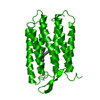
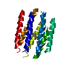
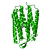
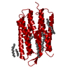
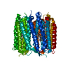
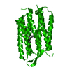
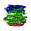
Mass: 24990.535 Da / Num. of mol.: 1 / Source method: isolated from a natural source Details: CONFORMER B IS GROUND STATE, CONFORMER A IS THE L-STATE FORM. Source: (natural) HALOBACTERIUM SALINARIUM (Halophile) / Strain: S9 / References: UniProt: P02945
Type: MARRESEARCH / Detector: CCD / Date: Oct 15, 1999
Radiation
Protocol: SINGLE WAVELENGTH / Monochromatic (M) / Laue (L): M / Scattering type: x-ray
Radiation wavelength
Wavelength: 0.934 Å / Relative weight: 1
Reflection
Resolution: 2.1→30.5 Å / Num. obs: 13088 / % possible obs: 96.6 % / Redundancy: 5.6 % / Biso Wilson estimate: 19.4 Å2 / Rsym value: 0.063 / Net I/σ(I): 9
Reflection shell
Resolution: 2.1→2.21 Å / Redundancy: 5.8 % / Mean I/σ(I) obs: 1.4 / Rsym value: 0.521 / % possible all: 99.2
Reflection
*PLUS
Num. measured all: 73411 / Rmerge(I) obs: 0.063
Reflection shell
*PLUS
% possible obs: 99.2 % / Rmerge(I) obs: 0.521
-
Processing
Software
Name
Version
Classification
CNS
1
refinement
DENZO
datareduction
SCALA
datascaling
SCALEPACK
datascaling
CNS
1
phasing
Refinement
Method to determine structure: OTHER / Resolution: 2.1→30.5 Å / Rfactor Rfree error: 0.012 / Data cutoff high absF: 2443186 / Isotropic thermal model: RESTRAINED / Cross valid method: THROUGHOUT / σ(F): 0 Details: THE COORDINATE FILE CONTAINS 2 MODELS CHAINS A AND B, ONLY A WAS REFINED, B WAS FIXED DURING THE WHOLE REFINEMENT. THE NUMBER OF ATOMS USED IN THE REFINEMENT REFERES TO CHAIN A. FREE R VALUE ...Details: THE COORDINATE FILE CONTAINS 2 MODELS CHAINS A AND B, ONLY A WAS REFINED, B WAS FIXED DURING THE WHOLE REFINEMENT. THE NUMBER OF ATOMS USED IN THE REFINEMENT REFERES TO CHAIN A. FREE R VALUE TEST SET SAME AS PREVIOUS REFINEMENTS 1QHJ, 1QKP, 1QKO
In the structure databanks used in Yorodumi, some data are registered as the other names, "COVID-19 virus" and "2019-nCoV". Here are the details of the virus and the list of structure data.
Jan 31, 2019. EMDB accession codes are about to change! (news from PDBe EMDB page)
EMDB accession codes are about to change! (news from PDBe EMDB page)
The allocation of 4 digits for EMDB accession codes will soon come to an end. Whilst these codes will remain in use, new EMDB accession codes will include an additional digit and will expand incrementally as the available range of codes is exhausted. The current 4-digit format prefixed with “EMD-” (i.e. EMD-XXXX) will advance to a 5-digit format (i.e. EMD-XXXXX), and so on. It is currently estimated that the 4-digit codes will be depleted around Spring 2019, at which point the 5-digit format will come into force.
The EM Navigator/Yorodumi systems omit the EMD- prefix.
Related info.:Q: What is EMD? / ID/Accession-code notation in Yorodumi/EM Navigator
Yorodumi is a browser for structure data from EMDB, PDB, SASBDB, etc.
This page is also the successor to EM Navigator detail page, and also detail information page/front-end page for Omokage search.
The word "yorodu" (or yorozu) is an old Japanese word meaning "ten thousand". "mi" (miru) is to see.
Related info.:EMDB / PDB / SASBDB / Comparison of 3 databanks / Yorodumi Search / Aug 31, 2016. New EM Navigator & Yorodumi / Yorodumi Papers / Jmol/JSmol / Function and homology information / Changes in new EM Navigator and Yorodumi
 Movie
Movie Controller


 Open data
 Basic information
 Components
 Keywords
 Function and homology information
 HALOBACTERIUM SALINARIUM (Halophile)
 X-RAY DIFFRACTION /
  Authors
 Citation
 Structure visualization
 Downloads & links
 Other downloads


 PDBj
PDBj


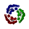
 Assembly
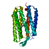

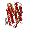
 Mass: 284.436 Da / Num. of mol.: 1 / Source method: obtained synthetically / Formula: C20H28O
 Mass: 18.015 Da / Num. of mol.: 30 / Source method: isolated from a natural source / Formula: H2O
 Sample preparation
 / Beamline: ID14-1 / Wavelength: 0.934
  Processing