 Movie
Movie Controller

[English] 日本語
 Yorodumi
- PDB-1qhj: X-RAY STRUCTURE OF BACTERIORHODOPSIN GROWN IN LIPIDIC CUBIC PHASES -
+ Open data

- Basic information
| Entry | Database: PDB / ID: 1qhj | ||||||
|---|---|---|---|---|---|---|---|
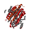
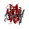
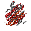
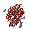
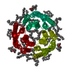
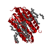
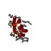
| Title | X-RAY STRUCTURE OF BACTERIORHODOPSIN GROWN IN LIPIDIC CUBIC PHASES | ||||||
 Components Components | PROTEIN (BACTERIORHODOPSIN) | ||||||
 Keywords Keywords | PHOTORECEPTOR / PROTON PUMP / MEMBRANE PROTEIN / RETINAL PROTEIN / LIPIDIC CUBIC PHASES / PURPLE MEMBRANE / ARCHEAL LIPIDS | ||||||
| Function / homology |  Function and homology information Function and homology informationlight-driven active monoatomic ion transmembrane transporter activity / photoreceptor activity / phototransduction / monoatomic ion channel activity / proton transmembrane transport / plasma membrane Similarity search - Function | ||||||
| Biological species |  Halobacterium salinarum (Halophile) Halobacterium salinarum (Halophile) | ||||||
| Method |  X-RAY DIFFRACTION / SYNCHROTRON / MOLECULAR REPLACEMENT / Resolution: 1.9 Å X-RAY DIFFRACTION / SYNCHROTRON / MOLECULAR REPLACEMENT / Resolution: 1.9 Å | ||||||
 Authors Authors | Belrhali, H. / Nollert, P. / Royant, A. / Menzel, C. / Rosenbusch, J.P. / Landau, E.M. / Pebay-Peyroula, E. | ||||||
 Citation Citation | Journal: Structure Fold.Des. / Year: 1999 Title: Protein, lipid and water organization in bacteriorhodopsin crystals: a molecular view of the purple membrane at 1.9 A resolution. Authors: Belrhali, H. / Nollert, P. / Royant, A. / Menzel, C. / Rosenbusch, J.P. / Landau, E.M. / Pebay-Peyroula, E. | ||||||
| History |
|
- Structure visualization
| Structure viewer | Molecule: MolmilJmol/JSmol |
|---|
- Downloads & links
-Download
| PDBx/mmCIF format | 1qhj.cif.gz | 67.1 KB | Display | PDBx/mmCIF format |
|---|---|---|---|---|
| PDB format | pdb1qhj.ent.gz | 50.9 KB | Display | PDB format |
| PDBx/mmJSON format | 1qhj.json.gz | Tree view | PDBx/mmJSON format | |
| Others |  Other downloads Other downloads |
-Validation report
| Arichive directory | https://data.pdbj.org/pub/pdb/validation_reports/qh/1qhjftp://data.pdbj.org/pub/pdb/validation_reports/qh/1qhj | HTTPS FTP |
|---|
-Related structure data
| Related structure data |  1ap9S S: Starting model for refinement |
|---|---|
| Similar structure data |

-Links
 PDBj
PDBj


- Assembly
| Deposited unit | 
| ||||||||
|---|---|---|---|---|---|---|---|---|---|
| 1 | 
| ||||||||
| Unit cell |
|
-Components
| #1: Protein | Mass: 26814.412 Da / Num. of mol.: 1 / Source method: isolated from a natural source / Details: RETINAL LINKED TO LYS 216 VIA A SCHIFF BASE / Source: (natural) Halobacterium salinarum (Halophile) / Cellular location: PLASMA MEMBRANE / Strain: S9 / References: UniProt: P02945 | ||||
|---|---|---|---|---|---|
| #2: Chemical | ChemComp-RET /   Mass: 284.436 Da / Num. of mol.: 1 / Source method: obtained synthetically / Formula: C20H28O Mass: 284.436 Da / Num. of mol.: 1 / Source method: obtained synthetically / Formula: C20H28O | ||||
| #3: Chemical | ChemComp-PH1 /   Mass: 637.158 Da / Num. of mol.: 9 / Source method: obtained synthetically / Formula: C43H88O2 Mass: 637.158 Da / Num. of mol.: 9 / Source method: obtained synthetically / Formula: C43H88O2#4: Water | ChemComp-HOH / |  Mass: 18.015 Da / Num. of mol.: 26 / Source method: isolated from a natural source / Formula: H2O Mass: 18.015 Da / Num. of mol.: 26 / Source method: isolated from a natural source / Formula: H2OHas protein modification | Y | |
-Experimental details
-Experiment
| Experiment | Method: X-RAY DIFFRACTION / Number of used crystals: 1 |
|---|
- Sample preparation
| Crystal | Density Matthews: 2.39 Å3/Da / Density % sol: 48 % | |||||||||||||||||||||||||
|---|---|---|---|---|---|---|---|---|---|---|---|---|---|---|---|---|---|---|---|---|---|---|---|---|---|---|
| Crystal grow | pH: 5.6 Details: PROTEIN FROM THE PURPLE MEMBRANE WAS DELIPIDATED AND RESOLVED IN OCTYL GLUCOSIDE. PROTEIN WAS CRYSTALLIZED FROM 60 - 70% (W/W) MONOOLEIN, 0.7 - 4.0 M NA/K - PHOSPHATE IN A PHOSPHATE BUFFER ...Details: PROTEIN FROM THE PURPLE MEMBRANE WAS DELIPIDATED AND RESOLVED IN OCTYL GLUCOSIDE. PROTEIN WAS CRYSTALLIZED FROM 60 - 70% (W/W) MONOOLEIN, 0.7 - 4.0 M NA/K - PHOSPHATE IN A PHOSPHATE BUFFER AT PH 5.6, AT 20C AND IN THE DARK. THE MIXTURE WAS CENTRIFUGED AT 10000G FOR 150 MN PRIOR TO CRYSTALLISATION. | |||||||||||||||||||||||||
| Crystal | *PLUS | |||||||||||||||||||||||||
| Crystal grow | *PLUS Temperature: 20 ℃ / Method: unknownDetails: Landau, E.M., (1996) Proc.Natl.Acad.Sci.USA., 93, 14532. | |||||||||||||||||||||||||
| Components of the solutions | *PLUS
|
-Data collection
| Diffraction | Mean temperature: 100 K |
|---|---|
| Diffraction source | Source: SYNCHROTRON / Site: ESRF  / Beamline: ID14-3 / Wavelength: 0.93 / Beamline: ID14-3 / Wavelength: 0.93 |
| Detector | Type: MARRESEARCH / Detector: CCD / Date: Dec 1, 1998 |
| Radiation | Protocol: SINGLE WAVELENGTH / Monochromatic (M) / Laue (L): M / Scattering type: x-ray |
| Radiation wavelength | Wavelength: 0.93 Å / Relative weight: 1 |
| Reflection | Resolution: 1.9→38 Å / Num. obs: 17996 / % possible obs: 99.5 % / Observed criterion σ(I): 0 / Redundancy: 5.4 % / Biso Wilson estimate: 29.3 Å2 / Rsym value: 0.046 / Net I/σ(I): 9.9 |
| Reflection shell | Resolution: 1.9→2 Å / Redundancy: 4 % / Mean I/σ(I) obs: 2.6 / Rsym value: 0.287 / % possible all: 98.3 |
| Reflection | *PLUS Num. measured all: 97807 / Rmerge(I) obs: 0.046 |
| Reflection shell | *PLUS Highest resolution: 1.9 Å / % possible obs: 98.3 % / Rmerge(I) obs: 0.287 |
- Processing
| Software |
| ||||||||||||||||||||||||||||||||||||||||||||||||||||||||||||
|---|---|---|---|---|---|---|---|---|---|---|---|---|---|---|---|---|---|---|---|---|---|---|---|---|---|---|---|---|---|---|---|---|---|---|---|---|---|---|---|---|---|---|---|---|---|---|---|---|---|---|---|---|---|---|---|---|---|---|---|---|---|
| Refinement | Method to determine structure: MOLECULAR REPLACEMENT Starting model: PDB ENTRY 1AP9 Resolution: 1.9→38 Å / Data cutoff high rms absF: 10000000 / Cross valid method: THROUGHOUT / σ(F): 0 Details: THE DATA USED FOR THIS REFINEMENT WERE COLLECTED FROM A NON-TWINNED CRYSTAL. PROTEIN, RETINAL AND WATER ATOMS WERE REFINED. NINE PHYTANYL MOIETIES COULD THEN BE MODELED IN THE ELECTRON ...Details: THE DATA USED FOR THIS REFINEMENT WERE COLLECTED FROM A NON-TWINNED CRYSTAL. PROTEIN, RETINAL AND WATER ATOMS WERE REFINED. NINE PHYTANYL MOIETIES COULD THEN BE MODELED IN THE ELECTRON DENSITY MAPS. THE REFINEMENT STATISTICS GIVEN BELOW CORRESPOND TO THE REFINEMENT WITHOUT LIPID MOLECULES.
| ||||||||||||||||||||||||||||||||||||||||||||||||||||||||||||
| Solvent computation | Solvent model: FLAT MODEL / Bsol: 83 Å2 / ksol: 0.435 e/Å3 | ||||||||||||||||||||||||||||||||||||||||||||||||||||||||||||
| Displacement parameters | Biso mean: 33.2 Å2
| ||||||||||||||||||||||||||||||||||||||||||||||||||||||||||||
| Refine analyze |
| ||||||||||||||||||||||||||||||||||||||||||||||||||||||||||||
| Refinement step | Cycle: LAST / Resolution: 1.9→38 Å
| ||||||||||||||||||||||||||||||||||||||||||||||||||||||||||||
| Refine LS restraints |
| ||||||||||||||||||||||||||||||||||||||||||||||||||||||||||||
| LS refinement shell | Resolution: 1.9→2 Å / Total num. of bins used: 8
| ||||||||||||||||||||||||||||||||||||||||||||||||||||||||||||
| Xplor file |
| ||||||||||||||||||||||||||||||||||||||||||||||||||||||||||||
| Software | *PLUS Name: CNS / Version: 5 / Classification: refinement | ||||||||||||||||||||||||||||||||||||||||||||||||||||||||||||
| Refinement | *PLUS Rfactor obs: 0.224 | ||||||||||||||||||||||||||||||||||||||||||||||||||||||||||||
| Solvent computation | *PLUS | ||||||||||||||||||||||||||||||||||||||||||||||||||||||||||||
| Displacement parameters | *PLUS Biso mean: 33.2 Å2 | ||||||||||||||||||||||||||||||||||||||||||||||||||||||||||||
| Refine LS restraints | *PLUS
| ||||||||||||||||||||||||||||||||||||||||||||||||||||||||||||
| LS refinement shell | *PLUS Highest resolution: 1.9 Å / Lowest resolution: 2 Å / % reflection Rfree: 5 % |
