 Movie
Movie Controller


+ Open data

- Basic information
| Entry | Database: PDB / ID: 2i1x | ||||||
|---|---|---|---|---|---|---|---|
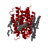
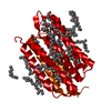
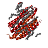
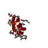
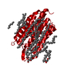
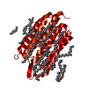
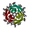
| Title | Bacteriorhodopsin/lipid complex, D96A mutant | ||||||
 Components Components | Bacteriorhodopsin | ||||||
 Keywords Keywords | TRANSPORT PROTEIN / ION PUMP / MEMBRANE PROTEIN / RETINAL PROTEIN / LIPIDS / PHOTORECEPTOR / HALOARCHAEA / 7-TRANSMEMBRANE / SERPENTINE / ION TRANSPORT | ||||||
| Function / homology |  Function and homology information Function and homology informationlight-driven active monoatomic ion transmembrane transporter activity / photoreceptor activity / phototransduction / monoatomic ion channel activity / proton transmembrane transport / plasma membrane Similarity search - Function | ||||||
| Biological species |  Halobacterium salinarum (Halophile) Halobacterium salinarum (Halophile) | ||||||
| Method |  X-RAY DIFFRACTION / SYNCHROTRON / MOLECULAR REPLACEMENT / Resolution: 2 Å X-RAY DIFFRACTION / SYNCHROTRON / MOLECULAR REPLACEMENT / Resolution: 2 Å | ||||||
 Authors Authors | Lanyi, J.K. / Schobert, B. | ||||||
 Citation Citation | Journal: Biochemistry / Year: 2006 Title: Propagating Structural Perturbation Inside Bacteriorhodopsin: Crystal Structures of the M State and the D96A and T46V Mutants. Authors: Lanyi, J.K. / Schobert, B. | ||||||
| History |
| ||||||
| Remark 600 | SCHIFF BASE LINKAGE BETWEEN LYS 216 (NZ) AND RET 301 (C15) DIETHER LIPID BILAYER |
- Structure visualization
| Structure viewer | Molecule: MolmilJmol/JSmol |
|---|
- Downloads & links
-Download
| PDBx/mmCIF format | 2i1x.cif.gz | 69.4 KB | Display | PDBx/mmCIF format |
|---|---|---|---|---|
| PDB format | pdb2i1x.ent.gz | 49.1 KB | Display | PDB format |
| PDBx/mmJSON format | 2i1x.json.gz | Tree view | PDBx/mmJSON format | |
| Others |  Other downloads Other downloads |
-Validation report
| Arichive directory | https://data.pdbj.org/pub/pdb/validation_reports/i1/2i1xftp://data.pdbj.org/pub/pdb/validation_reports/i1/2i1x | HTTPS FTP |
|---|
-Related structure data
| Related structure data |  2i20C  2i21C  1c3wS S: Starting model for refinement C: citing same article ( |
|---|---|
| Similar structure data |


-Links
 PDBj
PDBj


- Assembly
| Deposited unit | 
| ||||||||
|---|---|---|---|---|---|---|---|---|---|
| 1 | 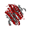
| ||||||||
| Unit cell |
|
-Components
| #1: Protein | Mass: 26885.490 Da / Num. of mol.: 1 / Mutation: D96A Source method: isolated from a genetically manipulated source Source: (gene. exp.) Halobacterium salinarum (Halophile) / Cellular location: PLASMA MEMBRANE / Plasmid: PBA2 / Cellular location (production host): CYTOPLASM / Production host: Halobacterium salinarum (Halophile) / Strain (production host): MPK409 / References: UniProt: P02945 | ||||||
|---|---|---|---|---|---|---|---|
| #2: Chemical | ChemComp-RET /   Mass: 284.436 Da / Num. of mol.: 1 / Source method: obtained synthetically / Formula: C20H28O Mass: 284.436 Da / Num. of mol.: 1 / Source method: obtained synthetically / Formula: C20H28O | ||||||
| #3: Chemical | ChemComp-LI1 /   Mass: 639.130 Da / Num. of mol.: 13 / Source method: obtained synthetically / Formula: C42H86O3 Mass: 639.130 Da / Num. of mol.: 13 / Source method: obtained synthetically / Formula: C42H86O3#4: Chemical | ChemComp-SQU / |   Mass: 380.734 Da / Num. of mol.: 1 / Source method: obtained synthetically / Formula: C27H56 Mass: 380.734 Da / Num. of mol.: 1 / Source method: obtained synthetically / Formula: C27H56#5: Water | ChemComp-HOH / |  Mass: 18.015 Da / Num. of mol.: 23 / Source method: isolated from a natural source / Formula: H2O Mass: 18.015 Da / Num. of mol.: 23 / Source method: isolated from a natural source / Formula: H2OHas protein modification | Y | |
-Experimental details
-Experiment
| Experiment | Method: X-RAY DIFFRACTION / Number of used crystals: 1 |
|---|
- Sample preparation
| Crystal | Density Matthews: 1.8 Å3/Da / Density % sol: 47.91 % |
|---|---|
| Crystal grow | Temperature: 295 K / Method: cubic lipid phase / pH: 5.6 Details: MO:WATER:PHOSPHATE, pH 5.60, CUBIC LIPID PHASE, temperature 295K |
-Data collection
| Diffraction | Mean temperature: 100 K |
|---|---|
| Diffraction source | Source: SYNCHROTRON / Site: SSRL  / Beamline: BL9-1 / Wavelength: 0.97977 / Wavelength: 0.97977 Å / Beamline: BL9-1 / Wavelength: 0.97977 / Wavelength: 0.97977 Å |
| Detector | Type: ADSC QUANTUM / Detector: CDD / Date: Apr 14, 2005 |
| Radiation | Protocol: SINGLE WAVELENGTH / Monochromatic (M) / Laue (L): M / Scattering type: x-ray |
| Radiation wavelength | Wavelength: 0.97977 Å / Relative weight: 1 |
| Reflection | Resolution: 2→25 Å / Num. obs: 15618 / % possible obs: 99.99 % / Observed criterion σ(F): 2 / Observed criterion σ(I): 2 / Rmerge(I) obs: 0.062 / Net I/σ(I): 23.4 |
| Reflection shell | Resolution: 2→2.09 Å / Rmerge(I) obs: 0.756 / Mean I/σ(I) obs: 2.9 / Num. unique all: 15618 / % possible all: 99.9 |
- Processing
| Software |
| |||||||||||||||||||||||||||||||||
|---|---|---|---|---|---|---|---|---|---|---|---|---|---|---|---|---|---|---|---|---|---|---|---|---|---|---|---|---|---|---|---|---|---|---|
| Refinement | Method to determine structure: MOLECULAR REPLACEMENT Starting model: PDB ENTRY 1C3W Resolution: 2→25 Å / Num. parameters: 8284 / Num. restraintsaints: 8217 / Cross valid method: FREE R / σ(F): 0 / Stereochemistry target values: ENGH AND HUBER / Details: NO MEROHEDRAL TWINNING
| |||||||||||||||||||||||||||||||||
| Refine analyze | Num. disordered residues: 0 / Occupancy sum non hydrogen: 2067 | |||||||||||||||||||||||||||||||||
| Refinement step | Cycle: LAST / Resolution: 2→25 Å
| |||||||||||||||||||||||||||||||||
| Refine LS restraints |
|
