 Movie
Movie Controller

[English] 日本語
 Yorodumi
- PDB-1ap9: X-RAY STRUCTURE OF BACTERIORHODOPSIN FROM MICROCRYSTALS GROWN IN ... -
+ Open data

- Basic information
| Entry | Database: PDB / ID: 1ap9 | ||||||
|---|---|---|---|---|---|---|---|
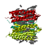
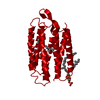
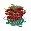
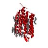
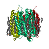
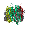
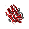
| Title | X-RAY STRUCTURE OF BACTERIORHODOPSIN FROM MICROCRYSTALS GROWN IN LIPIDIC CUBIC PHASES | ||||||
 Components Components | BACTERIORHODOPSIN | ||||||
 Keywords Keywords | PHOTORECEPTOR / PROTON PUMP / MEMBRANE PROTEIN / RETINAL PROTEIN / MICROCRYSTALS / MICROFOCUS BEAM / LIPIDIC CUBIC PHASES | ||||||
| Function / homology |  Function and homology information Function and homology informationlight-driven active monoatomic ion transmembrane transporter activity / photoreceptor activity / phototransduction / monoatomic ion channel activity / proton transmembrane transport / plasma membrane Similarity search - Function | ||||||
| Biological species |  Halobacterium salinarum (Halophile) Halobacterium salinarum (Halophile) | ||||||
| Method |  X-RAY DIFFRACTION / SYNCHROTRON / MOLECULAR REPLACEMENT / Resolution: 2.35 Å X-RAY DIFFRACTION / SYNCHROTRON / MOLECULAR REPLACEMENT / Resolution: 2.35 Å | ||||||
 Authors Authors | Pebay-Peyroula, E. / Rummel, G. / Rosenbusch, J.P. / Landau, E.M. | ||||||
 Citation Citation | Journal: Science / Year: 1997 Title: X-ray structure of bacteriorhodopsin at 2.5 angstroms from microcrystals grown in lipidic cubic phases. Authors: Pebay-Peyroula, E. / Rummel, G. / Rosenbusch, J.P. / Landau, E.M. #1: Journal: J.Phys.Chem.B / Year: 1997Title: Crystallisation of a Polar Protein and Small Molecules from the Aqueous Compartment of Lipidic Cubic Phases Authors: Landau, E.M. / Rummel, G. / Cowan-Jacob, S.W. / Rosenbusch, J.P. | ||||||
| History |
|
- Structure visualization
| Structure viewer | Molecule: MolmilJmol/JSmol |
|---|
- Downloads & links
-Download
| PDBx/mmCIF format | 1ap9.cif.gz | 56.3 KB | Display | PDBx/mmCIF format |
|---|---|---|---|---|
| PDB format | pdb1ap9.ent.gz | 40.9 KB | Display | PDB format |
| PDBx/mmJSON format | 1ap9.json.gz | Tree view | PDBx/mmJSON format | |
| Others |  Other downloads Other downloads |
-Validation report
| Arichive directory | https://data.pdbj.org/pub/pdb/validation_reports/ap/1ap9ftp://data.pdbj.org/pub/pdb/validation_reports/ap/1ap9 | HTTPS FTP |
|---|
-Related structure data
| Related structure data |  2brdS S: Starting model for refinement |
|---|---|
| Similar structure data |


-Links
 PDBj
PDBj


- Assembly
| Deposited unit | 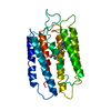
| ||||||||
|---|---|---|---|---|---|---|---|---|---|
| 1 | 
| ||||||||
| Unit cell |
|
-Components
| #1: Protein | Mass: 26814.412 Da / Num. of mol.: 1 / Source method: isolated from a natural source / Details: RETINAL LINKED TO LYS 216 VIA A SCHIFF BASE / Source: (natural) Halobacterium salinarum (Halophile) / Cellular location: PLASMA MEMBRANE / Strain: S9 / References: UniProt: P02945 |
|---|---|
| #2: Chemical | ChemComp-RET /   Mass: 284.436 Da / Num. of mol.: 1 / Source method: obtained synthetically / Formula: C20H28O Mass: 284.436 Da / Num. of mol.: 1 / Source method: obtained synthetically / Formula: C20H28O |
| #3: Water | ChemComp-HOH /  Mass: 18.015 Da / Num. of mol.: 26 / Source method: isolated from a natural source / Formula: H2O Mass: 18.015 Da / Num. of mol.: 26 / Source method: isolated from a natural source / Formula: H2O |
| Has protein modification | Y |
-Experimental details
-Experiment
| Experiment | Method: X-RAY DIFFRACTION / Number of used crystals: 1 |
|---|
- Sample preparation
| Crystal | Density Matthews: 2.39 Å3/Da / Density % sol: 48 % | |||||||||||||||||||||||||
|---|---|---|---|---|---|---|---|---|---|---|---|---|---|---|---|---|---|---|---|---|---|---|---|---|---|---|
| Crystal grow | pH: 5.6 Details: PROTEIN FROM THE PURPLE MEMBRANE WAS DELIPIDATED AND SOLUBILIZED IN OCTYL GLUCOSIDE. PROTEIN WAS CRYSTALLIZED FROM 60 - 70% (W/W) MONOOLEIN, 0.7 - 4.0 M NA/K - PHOSPHATE IN A PHOSPHATE ...Details: PROTEIN FROM THE PURPLE MEMBRANE WAS DELIPIDATED AND SOLUBILIZED IN OCTYL GLUCOSIDE. PROTEIN WAS CRYSTALLIZED FROM 60 - 70% (W/W) MONOOLEIN, 0.7 - 4.0 M NA/K - PHOSPHATE IN A PHOSPHATE BUFFER AT PH 5.6, AT 20C AND IN THE DARK. THE MIXTURE WAS CENTRIFUGED AT 10000G FOR 150 MN PRIOR TO CRYSTALLISATION. | |||||||||||||||||||||||||
| Crystal | *PLUS | |||||||||||||||||||||||||
| Crystal grow | *PLUS Temperature: 20 ℃ / Method: unknownDetails: Landau, E.M., (1996) Proc.Natl.Acad.Sci.USA., 93, 14532. | |||||||||||||||||||||||||
| Components of the solutions | *PLUS
|
-Data collection
| Diffraction | Mean temperature: 100 K |
|---|---|
| Diffraction source | Source: SYNCHROTRON / Site: ESRF  / Beamline: ID13 / Wavelength: 0.688 / Beamline: ID13 / Wavelength: 0.688 |
| Detector | Type: MARRESEARCH / Detector: IMAGE PLATE / Date: Aug 1, 1996 / Details: ELLIPSOIDAL MIRROR |
| Radiation | Monochromator: SI(111) / Monochromatic (M) / Laue (L): M / Scattering type: x-ray |
| Radiation wavelength | Wavelength: 0.688 Å / Relative weight: 1 |
| Reflection | Resolution: 2.35→10 Å / Num. obs: 8045 / % possible obs: 91.2 % / Observed criterion σ(I): 0 / Redundancy: 2 % / Biso Wilson estimate: 45.5 Å2 / Rsym value: 0.1 / Net I/σ(I): 4.7 |
| Reflection shell | Resolution: 2.35→2.56 Å / Redundancy: 1.6 % / Mean I/σ(I) obs: 2.4 / Rsym value: 0.266 / % possible all: 90.4 |
| Reflection | *PLUS Rmerge(I) obs: 0.1 |
- Processing
| Software |
| ||||||||||||||||||||||||||||||||||||||||||||||||||||||||||||
|---|---|---|---|---|---|---|---|---|---|---|---|---|---|---|---|---|---|---|---|---|---|---|---|---|---|---|---|---|---|---|---|---|---|---|---|---|---|---|---|---|---|---|---|---|---|---|---|---|---|---|---|---|---|---|---|---|---|---|---|---|---|
| Refinement | Method to determine structure: MOLECULAR REPLACEMENT Starting model: PDB ENTRY 2BRD Resolution: 2.35→5 Å / Data cutoff high absF: 10000000 / Data cutoff low absF: 0.001 / Cross valid method: THROUGHOUT / σ(F): 2.5
| ||||||||||||||||||||||||||||||||||||||||||||||||||||||||||||
| Displacement parameters | Biso mean: 75 Å2
| ||||||||||||||||||||||||||||||||||||||||||||||||||||||||||||
| Refine analyze |
| ||||||||||||||||||||||||||||||||||||||||||||||||||||||||||||
| Refinement step | Cycle: LAST / Resolution: 2.35→5 Å
| ||||||||||||||||||||||||||||||||||||||||||||||||||||||||||||
| Refine LS restraints |
| ||||||||||||||||||||||||||||||||||||||||||||||||||||||||||||
| LS refinement shell | Resolution: 2.35→2.48 Å / Total num. of bins used: 6
| ||||||||||||||||||||||||||||||||||||||||||||||||||||||||||||
| Xplor file |
| ||||||||||||||||||||||||||||||||||||||||||||||||||||||||||||
| Software | *PLUS Name: X-PLOR / Version: 3.8 / Classification: refinement | ||||||||||||||||||||||||||||||||||||||||||||||||||||||||||||
| Refinement | *PLUS Rfactor Rfree: 0.3179 | ||||||||||||||||||||||||||||||||||||||||||||||||||||||||||||
| Solvent computation | *PLUS | ||||||||||||||||||||||||||||||||||||||||||||||||||||||||||||
| Displacement parameters | *PLUS | ||||||||||||||||||||||||||||||||||||||||||||||||||||||||||||
| Refine LS restraints | *PLUS
| ||||||||||||||||||||||||||||||||||||||||||||||||||||||||||||
| LS refinement shell | *PLUS Rfactor Rfree: 0.384 |
