 Movie
Movie Controller

[English] 日本語
 Yorodumi
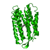
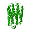
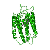
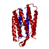
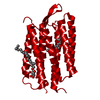
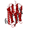
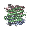
- PDB-1bm1: CRYSTAL STRUCTURE OF BACTERIORHODOPSIN IN THE LIGHT-ADAPTED STATE -
+ Open data

- Basic information
| Entry | Database: PDB / ID: 1bm1 | |||||||||
|---|---|---|---|---|---|---|---|---|---|---|
| Title | CRYSTAL STRUCTURE OF BACTERIORHODOPSIN IN THE LIGHT-ADAPTED STATE | |||||||||
 Components Components | BACTERIORHODOPSIN | |||||||||
 Keywords Keywords | PHOTORECEPTOR / PROTON PUMP / MEMBRANE PROTEIN / RETINAL PROTEIN | |||||||||
| Function / homology |  Function and homology information Function and homology informationlight-driven active monoatomic ion transmembrane transporter activity / photoreceptor activity / phototransduction / monoatomic ion channel activity / proton transmembrane transport / plasma membrane Similarity search - Function | |||||||||
| Biological species |  Halobacterium salinarum (Halophile) Halobacterium salinarum (Halophile) | |||||||||
| Method |  X-RAY DIFFRACTION / SYNCHROTRON / MOLECULAR REPLACEMENT / Resolution: 3.5 Å X-RAY DIFFRACTION / SYNCHROTRON / MOLECULAR REPLACEMENT / Resolution: 3.5 Å | |||||||||
 Authors Authors | Sato, H. / Takeda, K. / Tani, K. / Hino, T. / Okada, T. / Nakasako, M. / Kamiya, N. / Kouyama, T. | |||||||||
 Citation Citation | Journal: Acta Crystallogr.,Sect.D / Year: 1999 Title: Specific lipid-protein interactions in a novel honeycomb lattice structure of bacteriorhodopsin. Authors: Sato, H. / Takeda, K. / Tani, K. / Hino, T. / Okada, T. / Nakasako, M. / Kamiya, N. / Kouyama, T. #1: Journal: J.Mol.Biol. / Year: 1998Title: A Novel Three-Dimensional Crystal of Bacteriorhodopsin Obtained by Successive Fusion of the Vesicular Assemblies Authors: Takeda, K. / Sato, H. / Hino, T. / Kono, M. / Fukuda, K. / Sakurai, I. / Okada, T. / Kouyama, T. | |||||||||
| History |
|
- Structure visualization
| Structure viewer | Molecule: MolmilJmol/JSmol |
|---|
- Downloads & links
-Download
| PDBx/mmCIF format | 1bm1.cif.gz | 52.4 KB | Display | PDBx/mmCIF format |
|---|---|---|---|---|
| PDB format | pdb1bm1.ent.gz | 35.8 KB | Display | PDB format |
| PDBx/mmJSON format | 1bm1.json.gz | Tree view | PDBx/mmJSON format | |
| Others |  Other downloads Other downloads |
-Validation report
| Arichive directory | https://data.pdbj.org/pub/pdb/validation_reports/bm/1bm1ftp://data.pdbj.org/pub/pdb/validation_reports/bm/1bm1 | HTTPS FTP |
|---|
-Related structure data
| Related structure data |  2brdS S: Starting model for refinement |
|---|---|
| Similar structure data |

-Links
 PDBj
PDBj


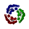
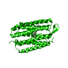
- Assembly
| Deposited unit | 
| ||||||||
|---|---|---|---|---|---|---|---|---|---|
| 1 | 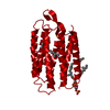
| ||||||||
| Unit cell |
|
-Components
| #1: Protein | Mass: 26797.381 Da / Num. of mol.: 1 / Source method: isolated from a natural source / Source: (natural) Halobacterium salinarum (Halophile) / Strain: JW3 / References: UniProt: P02945 |
|---|---|
| #2: Chemical | ChemComp-RET /   Mass: 284.436 Da / Num. of mol.: 1 / Source method: obtained synthetically / Formula: C20H28O Mass: 284.436 Da / Num. of mol.: 1 / Source method: obtained synthetically / Formula: C20H28O |
| #3: Chemical | ChemComp-DPG /   Mass: 887.195 Da / Num. of mol.: 1 / Source method: obtained synthetically / Formula: C46H96O11P2 / Comment: phospholipid*YM Mass: 887.195 Da / Num. of mol.: 1 / Source method: obtained synthetically / Formula: C46H96O11P2 / Comment: phospholipid*YM |
| Has protein modification | Y |
-Experimental details
-Experiment
| Experiment | Method: X-RAY DIFFRACTION / Number of used crystals: 1 |
|---|
- Sample preparation
| Crystal | Density Matthews: 3.37 Å3/Da / Density % sol: 63.47 % Description: DATA WERE COLLECTED USING THE WEISSENBERG METHOD | |||||||||||||||
|---|---|---|---|---|---|---|---|---|---|---|---|---|---|---|---|---|
| Crystal grow | pH: 5.2 / Details: pH 5.2 | |||||||||||||||
| Crystal grow | *PLUS Temperature: 278 K / Method: vapor diffusion | |||||||||||||||
| Components of the solutions | *PLUS
|
-Data collection
| Diffraction | Mean temperature: 285 K |
|---|---|
| Diffraction source | Source: SYNCHROTRON / Site: Photon Factory  / Beamline: BL-18B / Wavelength: 1.012 / Beamline: BL-18B / Wavelength: 1.012 |
| Detector | Type: FUJI / Detector: IMAGE PLATE / Date: Nov 14, 1996 |
| Radiation | Monochromatic (M) / Laue (L): M / Scattering type: x-ray |
| Radiation wavelength | Wavelength: 1.012 Å / Relative weight: 1 |
| Reflection | Resolution: 3.5→50 Å / Num. obs: 4000 / % possible obs: 80.2 % / Observed criterion σ(I): 2 / Redundancy: 7.4 % / Biso Wilson estimate: 22 Å2 / Rmerge(I) obs: 0.103 / Rsym value: 0.103 |
| Reflection shell | Resolution: 3.5→3.65 Å / Redundancy: 4.8 % / Rmerge(I) obs: 0.159 / Rsym value: 0.159 / % possible all: 45.2 |
| Reflection shell | *PLUS % possible obs: 45.2 % |
- Processing
| Software |
| ||||||||||||||||||||||||||||||||||||||||||||||||||||||||||||
|---|---|---|---|---|---|---|---|---|---|---|---|---|---|---|---|---|---|---|---|---|---|---|---|---|---|---|---|---|---|---|---|---|---|---|---|---|---|---|---|---|---|---|---|---|---|---|---|---|---|---|---|---|---|---|---|---|---|---|---|---|---|
| Refinement | Method to determine structure: MOLECULAR REPLACEMENT Starting model: PDB ENTRY 2BRD Resolution: 3.5→8 Å / σ(F): 2
| ||||||||||||||||||||||||||||||||||||||||||||||||||||||||||||
| Refinement step | Cycle: LAST / Resolution: 3.5→8 Å
| ||||||||||||||||||||||||||||||||||||||||||||||||||||||||||||
| Refine LS restraints |
| ||||||||||||||||||||||||||||||||||||||||||||||||||||||||||||
| Xplor file |
| ||||||||||||||||||||||||||||||||||||||||||||||||||||||||||||
| Software | *PLUS Name: X-PLOR / Version: 3.851 / Classification: refinement | ||||||||||||||||||||||||||||||||||||||||||||||||||||||||||||
| Refinement | *PLUS Rfactor obs: 0.269 | ||||||||||||||||||||||||||||||||||||||||||||||||||||||||||||
| Solvent computation | *PLUS | ||||||||||||||||||||||||||||||||||||||||||||||||||||||||||||
| Displacement parameters | *PLUS | ||||||||||||||||||||||||||||||||||||||||||||||||||||||||||||
| Refine LS restraints | *PLUS
| ||||||||||||||||||||||||||||||||||||||||||||||||||||||||||||
| LS refinement shell | *PLUS Rfactor Rfree: 0.361 / Num. reflection obs: 247 / Rfactor obs: 0.272 |
