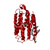
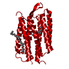
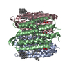
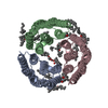
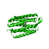
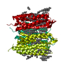
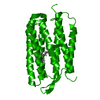
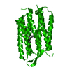
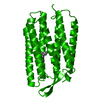
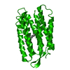
Journal: Nature / Year: 2000 Title: Molecular mechanism of vectorial proton translocation by bacteriorhodopsin. Authors: S Subramaniam / R Henderson / Abstract: Bacteriorhodopsin, a membrane protein with a relative molecular mass of 27,000, is a light driven pump which transports protons across the cell membrane of the halophilic organism Halobacterium ...Bacteriorhodopsin, a membrane protein with a relative molecular mass of 27,000, is a light driven pump which transports protons across the cell membrane of the halophilic organism Halobacterium salinarum. The chromophore retinal is covalently attached to the protein via a protonated Schiff base. Upon illumination, retinal is isomerized. The Schiff base then releases a proton to the extracellular medium, and is subsequently reprotonated from the cytoplasm. An atomic model for bacteriorhodopsin was first determined by Henderson et al, and has been confirmed and extended by work in a number of laboratories in the last few years. Here we present an atomic model for structural changes involved in the vectorial, light-driven transport of protons by bacteriorhodopsin. A 'switch' mechanism ensures the vectorial nature of pumping. First, retinal unbends, triggered by loss of the Schiff base proton, and second, a protein conformational change occurs. This conformational change, which we have determined by electron crystallography at atomic (3.2 A in-plane and 3.6 A vertical) resolution, is largely localized to helices F and G, and provides an 'opening' of the protein to protons on the cytoplasmic side of the membrane.
History
Deposition
Jul 15, 2000
Deposition site: RCSB / Processing site: RCSB
Revision 1.0
Aug 9, 2000
Provider: repository / Type: Initial release
Revision 1.1
Apr 27, 2008
Group: Version format compliance
Revision 1.2
Jul 13, 2011
Group: Derived calculations / Version format compliance
Mass: 284.436 Da / Num. of mol.: 1 / Source method: obtained synthetically / Formula: C20H28O
Has protein modification
Y
-
Experimental details
-
Experiment
Experiment
Method: ELECTRON CRYSTALLOGRAPHY / Number of used crystals: 402
EM experiment
Aggregation state: 2D ARRAY / 3D reconstruction method: electron crystallography
Crystal symmetry
∠γ: 120 ° / A: 62.45 Å / B: 62.45 Å / C: 100.9 Å / Space group name H-M: P3
-
Sample preparation
Component
Name: CYTOPLASMICALLY OPEN CONFORMATION OF BACTERIORHODOPSIN Source: RECOMBINANT
Molecular weight
Value: .027 MDa / Experimental value: NO
Source (natural)
Organism: Halobacterium salinarum (Halophile)
Specimen
Embedding applied: YES / Shadowing applied: NO / Staining applied: NO / Vitrification applied: YES
EM embedding
Material: glucose or trehalose
Crystal grow
Temperature: 310 K / Method: naturally occurring in vivo / pH: 7 Details: crystals are increased in size by fusion and annealing using detergents, pH 7, naturally occurring in vivo, temperature 37K
Crystal grow
*PLUS
Temperature: 4 ℃ / pH: 5.6 / Method: unknown
Components of the solutions
*PLUS
ID
Conc.
Common name
Crystal-ID
Sol-ID
1
18-23 mg/ml
protein
1
1
2
0.5 %(w/v)
beta-octylglucopyranoside
1
1
3
4 %(w/v)
benzamidine
1
1
4
1.75M
sodiumphosphate
1
1
5
1.8-2.3 M
ammoniumsulfate
1
reservoir
-
Data collection
Microscopy
Model: FEI/PHILIPS CM12 / Details: imaging date 1998-12-01
Electron gun
Illumination mode: FLOOD BEAM
Electron lens
Mode: DIFFRACTION
Image recording
Film or detector model: GENERIC CCD / Num. of diffraction images: 1000
Diffraction
Mean temperature: 93 K
Diffraction source
Source: ELECTRON MICROSCOPE / Type: OTHER / Wavelength: 0.033
Detector
Type: OTHER / Detector: CCD / Date: Dec 1, 1998
Radiation
Protocol: SINGLE WAVELENGTH / Monochromatic (M) / Laue (L): M / Scattering type: electron
∠γ: 120 ° / A: 62.45 Å / B: 62.45 Å / C: 100.9 Å / Space group name H-M: P3
3D reconstruction
Resolution: 3.2 Å / Resolution method: DIFFRACTION PATTERN/LAYERLINES Details: 402 patterns merged (merging R 17.3%) to generate set of lattice lines covering ~87% of reciprocal space, with resolution of 3.2 A in-plane and 3.6 A vertically [primary citation]
Refinement
Resolution: 3.2→200 Å / Stereochemistry target values: Engh & Huber Details: Each diffraction pattern was automatically indexed, and the spot intensities were integrated either using a raster (for patterns recorded at specimen tilts less than 30 degrees) or using ...Details: Each diffraction pattern was automatically indexed, and the spot intensities were integrated either using a raster (for patterns recorded at specimen tilts less than 30 degrees) or using profile fitting (for patterns recorded at specimen tilts at or greater than 30 degrees, which represented about 80% of the total data set). Each pattern was then compared to the curves recorded for wild-type bacteriorhodopsin in glucose at -100 degrees C [Ceska and Henderson J. Mol. Biol. 213: 539-560 (1990)], and the relative proportions of the four different twins determined. This exercise was carried out with all four theoretically possible orientations of the crystal axes relative to the previous reference curves to ensure that the data were merged correctly. From the initial set of 486 patterns chosen, 286 minimally twinned diffraction patterns were selected in which the major twin proportion was greater than 0.8. These 286 patterns were merged using the wild-type lattice lines as a reference and lattice lines were fitted to the data to obtain an initial approximately merged set of lattice lines (merge #1) describing the structure of the triple mutant. The original set of 486 patterns was then merged against the new lattice curves to redetermine the twin proportions more accurately. The merging parameters for each crystal were inspected carefully again, and 84 crystals for which the major twin proportion was less than 0.70 were excluded from the data set. The remaining 402 substantially untwinned diffraction patterns were used to generate a new set of curves and the procedure repeated to create a stable set of lattice lines. The merged data were further improved by using an estimate of sigma values for each reflection, and by the inclusion of an individual weighting factor for each diffraction pattern using procedures described by Grigorieff and Henderson [Ultramicroscopy 60: 295-309 (1995)]. Two cycles of this refinement were carried out to obtain a final set of merged curves. The curves were sampled at 1/100 Angstroms (approximately twice the thickness of the membrane) to obtain a set of intensities at H,K,L values so that the data could be further processed with standard X-ray crystallographic programs. For the tilt angles used, the maximal possible theoretical completeness of the data set is ~87%. The completeness of our data is close to this limit up to 3.5 Angstroms. The completeness drops to 77.7 % when all of the data to 3.2 Angstroms is included.
Rfactor
Num. reflection
% reflection
Selection details
Rfree
0.321
610
-
RANDOM
Rwork
0.272
-
-
-
all
-
7298
-
-
obs
-
5668
77.7 %
-
Refinement step
Cycle: LAST / Resolution: 3.2→200 Å
Protein
Nucleic acid
Ligand
Solvent
Total
Num. atoms
1726
0
20
0
1746
Refine LS restraints
Refine-ID
Type
Dev ideal
ELECTRONCRYSTALLOGRAPHY
c_bond_d
0.009
ELECTRONCRYSTALLOGRAPHY
c_angle_deg
1.4
+
About Yorodumi
-
News
-
Feb 9, 2022. New format data for meta-information of EMDB entries
New format data for meta-information of EMDB entries
Version 3 of the EMDB header file is now the official format.
The previous official version 1.9 will be removed from the archive.
In the structure databanks used in Yorodumi, some data are registered as the other names, "COVID-19 virus" and "2019-nCoV". Here are the details of the virus and the list of structure data.
Jan 31, 2019. EMDB accession codes are about to change! (news from PDBe EMDB page)
EMDB accession codes are about to change! (news from PDBe EMDB page)
The allocation of 4 digits for EMDB accession codes will soon come to an end. Whilst these codes will remain in use, new EMDB accession codes will include an additional digit and will expand incrementally as the available range of codes is exhausted. The current 4-digit format prefixed with “EMD-” (i.e. EMD-XXXX) will advance to a 5-digit format (i.e. EMD-XXXXX), and so on. It is currently estimated that the 4-digit codes will be depleted around Spring 2019, at which point the 5-digit format will come into force.
The EM Navigator/Yorodumi systems omit the EMD- prefix.
Related info.:Q: What is EMD? / ID/Accession-code notation in Yorodumi/EM Navigator
Yorodumi is a browser for structure data from EMDB, PDB, SASBDB, etc.
This page is also the successor to EM Navigator detail page, and also detail information page/front-end page for Omokage search.
The word "yorodu" (or yorozu) is an old Japanese word meaning "ten thousand". "mi" (miru) is to see.
Related info.:EMDB / PDB / SASBDB / Comparison of 3 databanks / Yorodumi Search / Aug 31, 2016. New EM Navigator & Yorodumi / Yorodumi Papers / Jmol/JSmol / Function and homology information / Changes in new EM Navigator and Yorodumi
 Movie
Movie Controller

 Yorodumi
 Open data
 Basic information
 Components
 Keywords
 Function and homology information
 Halobacterium salinarum (Halophile)
 Authors
 Citation
 Structure visualization
 Downloads & links
 Other downloads
 PDBj
PDBj


 Assembly
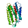

 Mass: 284.436 Da / Num. of mol.: 1 / Source method: obtained synthetically / Formula: C20H28O
 Sample preparation
 Processing