Mass: 18.015 Da / Num. of mol.: 10 / Source method: isolated from a natural source / Formula: H2O
Has protein modification
Y
-
Experimental details
-
Experiment
Experiment
Method: X-RAY DIFFRACTION / Number of used crystals: 1
-
Sample preparation
Crystal
Density Matthews: 2.49 Å3/Da / Density % sol: 50.61 % Description: thin plate microcrystals, 5-40 microns in the longest dimension
Crystal grow
Temperature: 294 K / Method: lipidic cubic phase / pH: 5.6 Details: Precipitant: 27%-39% PEG 2000, 100 mM phosphate buffer pH 5.6. MAG 9.9 used for LCP formation. Crystallization setup in Hamilton syringe with tube of LCP surrounded by precipitant. PH range: 5.4-5.8
-
Data collection
Diffraction
Mean temperature: 294 K Ambient temp details: ambient room temperature in the experimental hutch at ID13 ESRF
Resolution: 2.4→31.4 Å / Cor.coef. Fo:Fc: 0.945 / Cor.coef. Fo:Fc free: 0.921 / SU B: 23.607 / SU ML: 0.249 / Cross valid method: THROUGHOUT / σ(F): 0 / ESU R: 0.507 / ESU R Free: 0.272 / Stereochemistry target values: MAXIMUM LIKELIHOOD Details: HYDROGENS HAVE BEEN ADDED IN THE RIDING POSITIONS U VALUES : WITH TLS ADDED
Rfactor
Num. reflection
% reflection
Selection details
Rfree
0.249
441
4.6 %
RANDOM
Rwork
0.2053
9192
-
-
obs
0.2072
9633
99.96 %
-
Solvent computation
Ion probe radii: 0.8 Å / Shrinkage radii: 0.8 Å / VDW probe radii: 1.2 Å / Solvent model: MASK
In the structure databanks used in Yorodumi, some data are registered as the other names, "COVID-19 virus" and "2019-nCoV". Here are the details of the virus and the list of structure data.
Jan 31, 2019. EMDB accession codes are about to change! (news from PDBe EMDB page)
EMDB accession codes are about to change! (news from PDBe EMDB page)
The allocation of 4 digits for EMDB accession codes will soon come to an end. Whilst these codes will remain in use, new EMDB accession codes will include an additional digit and will expand incrementally as the available range of codes is exhausted. The current 4-digit format prefixed with “EMD-” (i.e. EMD-XXXX) will advance to a 5-digit format (i.e. EMD-XXXXX), and so on. It is currently estimated that the 4-digit codes will be depleted around Spring 2019, at which point the 5-digit format will come into force.
The EM Navigator/Yorodumi systems omit the EMD- prefix.
Related info.:Q: What is EMD? / ID/Accession-code notation in Yorodumi/EM Navigator
Yorodumi is a browser for structure data from EMDB, PDB, SASBDB, etc.
This page is also the successor to EM Navigator detail page, and also detail information page/front-end page for Omokage search.
The word "yorodu" (or yorozu) is an old Japanese word meaning "ten thousand". "mi" (miru) is to see.
Related info.:EMDB / PDB / SASBDB / Comparison of 3 databanks / Yorodumi Search / Aug 31, 2016. New EM Navigator & Yorodumi / Yorodumi Papers / Jmol/JSmol / Function and homology information / Changes in new EM Navigator and Yorodumi
 Movie
Movie Controller

 Yorodumi
 Open data
 Basic information
 Components
 Keywords
 Function and homology information
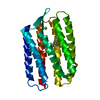
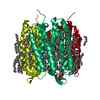
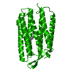
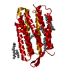
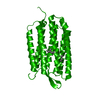
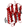
 Halobacterium salinarum (Halophile)
 X-RAY DIFFRACTION /
  Authors
 Citation
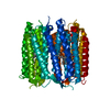
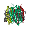
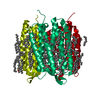
 Structure visualization
 Downloads & links
 Other downloads


 PDBj
PDBj


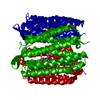
 Assembly

 Mass: 284.436 Da / Num. of mol.: 1 / Source method: obtained synthetically / Formula: C20H28O
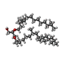
 Mass: 639.130 Da / Num. of mol.: 5 / Source method: obtained synthetically / Formula: C42H86O3
 Mass: 18.015 Da / Num. of mol.: 10 / Source method: isolated from a natural source / Formula: H2O
 Sample preparation
 / Beamline: ID13 / Wavelength: 1 Å
 Processing