 Movie
Movie Controller


+ Open data

- Basic information
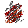
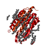
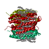
| Entry | Database: PDB / ID: 1kgb | ||||||
|---|---|---|---|---|---|---|---|
| Title | structure of ground-state bacteriorhodopsin | ||||||
 Components Components | bacteriorhodopsin | ||||||
 Keywords Keywords | PROTON TRANSPORT / bacteriorhodopsin / ground-state | ||||||
| Function / homology |  Function and homology information Function and homology informationlight-driven active monoatomic ion transmembrane transporter activity / photoreceptor activity / phototransduction / monoatomic ion channel activity / proton transmembrane transport / plasma membrane Similarity search - Function | ||||||
| Biological species |  Halobacterium salinarum (Halophile) Halobacterium salinarum (Halophile) | ||||||
| Method |  X-RAY DIFFRACTION / SYNCHROTRON / MOLECULAR REPLACEMENT / Resolution: 1.65 Å X-RAY DIFFRACTION / SYNCHROTRON / MOLECULAR REPLACEMENT / Resolution: 1.65 Å | ||||||
 Authors Authors | Facciotti, M.T. / Rouhani, S. / Burkard, F.T. / Betancourt, F.M. / Downing, K.H. / Rose, R.B. / McDermott, G. / Glaeser, R.M. | ||||||
 Citation Citation | Journal: Biophys.J. / Year: 2001 Title: Structure of an early intermediate in the M-state phase of the bacteriorhodopsin photocycle. Authors: Facciotti, M.T. / Rouhani, S. / Burkard, F.T. / Betancourt, F.M. / Downing, K.H. / Rose, R.B. / McDermott, G. / Glaeser, R.M. | ||||||
| History |
|
- Structure visualization
| Structure viewer | Molecule: MolmilJmol/JSmol |
|---|
- Downloads & links
-Download
| PDBx/mmCIF format | 1kgb.cif.gz | 71.3 KB | Display | PDBx/mmCIF format |
|---|---|---|---|---|
| PDB format | pdb1kgb.ent.gz | 51.1 KB | Display | PDB format |
| PDBx/mmJSON format | 1kgb.json.gz | Tree view | PDBx/mmJSON format | |
| Others |  Other downloads Other downloads |
-Validation report
| Arichive directory | https://data.pdbj.org/pub/pdb/validation_reports/kg/1kgbftp://data.pdbj.org/pub/pdb/validation_reports/kg/1kgb | HTTPS FTP |
|---|
-Related structure data
| Related structure data |  1kg8C  1kg9C 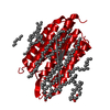 1c3wS S: Starting model for refinement C: citing same article ( |
|---|---|
| Similar structure data |


-Links
 PDBj
PDBj


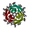
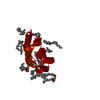
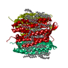
- Assembly
| Deposited unit | 
| ||||||||
|---|---|---|---|---|---|---|---|---|---|
| 1 | 
| ||||||||
| Unit cell |
|
-Components
| #1: Protein | Mass: 25301.916 Da / Num. of mol.: 1 / Source method: isolated from a natural source / Source: (natural) Halobacterium salinarum (Halophile) / References: UniProt: P02945 | ||||
|---|---|---|---|---|---|
| #2: Chemical | ChemComp-RET /   Mass: 284.436 Da / Num. of mol.: 1 / Source method: obtained synthetically / Formula: C20H28O Mass: 284.436 Da / Num. of mol.: 1 / Source method: obtained synthetically / Formula: C20H28O | ||||
| #3: Chemical | ChemComp-LI1 / 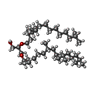  Mass: 639.130 Da / Num. of mol.: 13 / Source method: obtained synthetically / Formula: C42H86O3 Mass: 639.130 Da / Num. of mol.: 13 / Source method: obtained synthetically / Formula: C42H86O3#4: Water | ChemComp-HOH / |  Mass: 18.015 Da / Num. of mol.: 27 / Source method: isolated from a natural source / Formula: H2O Mass: 18.015 Da / Num. of mol.: 27 / Source method: isolated from a natural source / Formula: H2OHas protein modification | Y | |
-Experimental details
-Experiment
| Experiment | Method: X-RAY DIFFRACTION / Number of used crystals: 1 |
|---|
- Sample preparation
| Crystal | Density Matthews: 2.31 Å3/Da / Density % sol: 46.86 % | ||||||||||||||||||
|---|---|---|---|---|---|---|---|---|---|---|---|---|---|---|---|---|---|---|---|
| Crystal grow | Method: mono-olein cubic phase / pH: 5.6 Details: 3.0M Na/K Phosphate, pH 5.6, mono-olein cubic phase, temperature 100K | ||||||||||||||||||
| Crystal grow | *PLUS Method: unknown | ||||||||||||||||||
| Components of the solutions | *PLUS
|
-Data collection
| Diffraction | Mean temperature: 100 K |
|---|---|
| Diffraction source | Source: SYNCHROTRON / Site: ALS  / Beamline: 5.0.2 / Wavelength: 0.97 Å / Beamline: 5.0.2 / Wavelength: 0.97 Å |
| Detector | Type: ADSC QUANTUM 4 / Detector: CCD / Date: May 29, 1999 |
| Radiation | Monochromator: NULL / Protocol: SINGLE WAVELENGTH / Monochromatic (M) / Laue (L): M / Scattering type: x-ray |
| Radiation wavelength | Wavelength: 0.97 Å / Relative weight: 1 |
| Reflection | Resolution: 1.65→20 Å / Num. all: 26695 / Num. obs: 26695 / % possible obs: 98.7 % / Observed criterion σ(F): 0 / Observed criterion σ(I): 0 / Rmerge(I) obs: 0.036 / Net I/σ(I): 32.8 |
| Reflection shell | Resolution: 1.65→1.7 Å / Rmerge(I) obs: 0.358 / Mean I/σ(I) obs: 3.2 / % possible all: 96.8 |
| Reflection | *PLUS Lowest resolution: 20 Å / Rmerge(I) obs: 0.036 |
| Reflection shell | *PLUS % possible obs: 96.8 % / Rmerge(I) obs: 0.358 |
- Processing
| Software |
| ||||||||||||||||||||
|---|---|---|---|---|---|---|---|---|---|---|---|---|---|---|---|---|---|---|---|---|---|
| Refinement | Method to determine structure: MOLECULAR REPLACEMENT Starting model: 1C3W Resolution: 1.65→20 Å / σ(F): 0 / Stereochemistry target values: NULL
| ||||||||||||||||||||
| Refinement step | Cycle: LAST / Resolution: 1.65→20 Å
| ||||||||||||||||||||
| Refine LS restraints |
| ||||||||||||||||||||
| Software | *PLUS Name: SHELXL / Version: 97 / Classification: refinement | ||||||||||||||||||||
| Refinement | *PLUS Lowest resolution: 20 Å / Num. reflection obs: 25672 / σ(F): 0 / Rfactor all: 0.184 / Rfactor obs: 0.177 / Rfactor Rfree: 0.253 / Rfactor Rwork: 0.177 | ||||||||||||||||||||
| Solvent computation | *PLUS | ||||||||||||||||||||
| Displacement parameters | *PLUS | ||||||||||||||||||||
| Refine LS restraints | *PLUS
|
