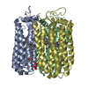
 Movie
Movie Controller

[English] 日本語
 Yorodumi
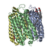
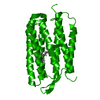
- PDB-1at9: STRUCTURE OF BACTERIORHODOPSIN AT 3.0 ANGSTROM DETERMINED BY ELEC... -
+ Open data

- Basic information
| Entry | Database: PDB / ID: 1at9 | |||||||||
|---|---|---|---|---|---|---|---|---|---|---|
| Title | STRUCTURE OF BACTERIORHODOPSIN AT 3.0 ANGSTROM DETERMINED BY ELECTRON CRYSTALLOGRAPHY | |||||||||
 Components Components | BACTERIORHODOPSIN | |||||||||
 Keywords Keywords | PHOTORECEPTOR / PROTON PUMP / MEMBRANE PROTEIN / RETINAL PROTEIN / TWO-DIMENSIONAL CRYSTAL | |||||||||
| Function / homology |  Function and homology information Function and homology informationlight-driven active monoatomic ion transmembrane transporter activity / photoreceptor activity / phototransduction / monoatomic ion channel activity / proton transmembrane transport / plasma membrane Similarity search - Function | |||||||||
| Biological species |  Halobacterium salinarum (Halophile) Halobacterium salinarum (Halophile) | |||||||||
| Method | ELECTRON CRYSTALLOGRAPHY / electron crystallography / cryo EM / Resolution: 2.8 Å | |||||||||
 Authors Authors | Kimura, Y. / Vassylyev, D.G. / Miyazawa, A. / Kidera, A. / Matsushima, M. / Mitsuoka, K. / Murata, K. / Hirai, T. / Fujiyoshi, Y. | |||||||||
 Citation Citation | Journal: Nature / Year: 1997 Title: Surface of bacteriorhodopsin revealed by high-resolution electron crystallography. Authors: Y Kimura / D G Vassylyev / A Miyazawa / A Kidera / M Matsushima / K Mitsuoka / K Murata / T Hirai / Y Fujiyoshi /  Abstract: Bacteriorhodopsin is a transmembrane protein that uses light energy, absorbed by its chromophore retinal, to pump protons from the cytoplasm of bacteria such as Halobacterium salinarium into the ...Bacteriorhodopsin is a transmembrane protein that uses light energy, absorbed by its chromophore retinal, to pump protons from the cytoplasm of bacteria such as Halobacterium salinarium into the extracellular space. It is made up of seven alpha-helices, and in the bacterium forms natural, two-dimensional crystals called purple membranes. We have analysed these crystals by electron cryo-microscopy to obtain images of bacteriorhodopsin at 3.0 A resolution. The structure covers nearly all 248 amino acids, including loops outside the membrane, and reveals the distribution of charged residues on both sides of the membrane surface. In addition, analysis of the electron-potential map produced by this method allows the determination of the charge status of these residues. On the extracellular side, four glutamate residues surround the entrance to the proton channel, whereas on the cytoplasmic side, four aspartic acids occur in a plane at the boundary of the hydrophobic-hydrophilic interface. The negative charges produced by these aspartate residues is encircled by areas of positive charge that may facilitate accumulation and lateral movement of protons on this surface. #1: Journal: J.Mol.Biol. / Year: 1996Title: Electron-Crystallographic Refinement of the Structure of Bacteriorhodopsin Authors: Grigorieff, N. / Ceska, T.A. / Downing, K.H. / Baldwin, J.M. / Henderson, R. #2: Journal: J.Mol.Biol. / Year: 1990Title: Model for the Structure of Bacteriorhodopsin Based on High-Resolution Electron Cryo-Microscopy Authors: Henderson, R. / Baldwin, J.M. / Ceska, T.A. / Zemlin, F. / Beckmann, E. / Downing, K.H. | |||||||||
| History |
|
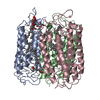
- Structure visualization
| Movie |
Movie viewer |
|---|---|
| Structure viewer | Molecule: MolmilJmol/JSmol |
- Downloads & links
-Download
| PDBx/mmCIF format | 1at9.cif.gz | 52.1 KB | Display | PDBx/mmCIF format |
|---|---|---|---|---|
| PDB format | pdb1at9.ent.gz | 35.4 KB | Display | PDB format |
| PDBx/mmJSON format | 1at9.json.gz | Tree view | PDBx/mmJSON format | |
| Others |  Other downloads Other downloads |
-Validation report
| Arichive directory | https://data.pdbj.org/pub/pdb/validation_reports/at/1at9ftp://data.pdbj.org/pub/pdb/validation_reports/at/1at9 | HTTPS FTP |
|---|
-Related structure data
| Similar structure data |
|---|


-Links
 PDBj
PDBj


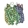
- Assembly
| Deposited unit | 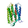
| ||||||||
|---|---|---|---|---|---|---|---|---|---|
| 1 | 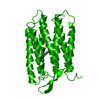
| ||||||||
| Unit cell |
|
-Components
| #1: Protein | Mass: 26797.381 Da / Num. of mol.: 1 / Source method: isolated from a natural source / Source: (natural) Halobacterium salinarum (Halophile) / Strain: JW5 / References: UniProt: P02945 |
|---|---|
| #2: Chemical | ChemComp-RET /   Mass: 284.436 Da / Num. of mol.: 1 / Source method: obtained synthetically / Formula: C20H28O Mass: 284.436 Da / Num. of mol.: 1 / Source method: obtained synthetically / Formula: C20H28O |
| Compound details | THE SURFACE STRUCTURE IS NEW AND UNIQUE. ALPHA CARBONS' POSITIONS ARE SIMILAR TO 2BRD EXCEPT FOR ...THE SURFACE STRUCTURE IS NEW AND UNIQUE. ALPHA CARBONS' POSITIONS ARE SIMILAR TO 2BRD EXCEPT FOR THOSE NEAR THE MEMBRANE SURFACES. SOME OF THE SIDE CHAINS DIFFER CONSIDERAB |
| Has protein modification | Y |
-Experimental details
-Experiment
| Experiment | Method: ELECTRON CRYSTALLOGRAPHY |
|---|---|
| EM experiment | Aggregation state: 2D ARRAY / 3D reconstruction method: electron crystallography |
- Sample preparation
| Component | Name: Bacteriorhodopsin / Type: COMPLEX / Source: NATURAL |
|---|---|
| Buffer solution | pH: 5.5 / Details: 0.4 M citric acid Na2HPO4, 3% trehalose |
| Specimen | Embedding applied: YES / Shadowing applied: NO / Staining applied: NO / Vitrification applied: YES |
| Specimen support | Details: specially ordered surface-polished to minimize wrinkles induced by cryo-fixation Grid material: MOLYBDENUM |
| EM embedding | Details: 3% trehalose / Material: trehalose |
| Vitrification | Instrument: REICHERT-JUNG PLUNGER / Cryogen name: ETHANE / Details: delay of 10 seconds before rapid freezing |
| Crystal | Density Matthews: 4.23 Å3/Da / Density % sol: 71 % |
| Crystal grow | *PLUS Method: other / Details: Oesterhelt, D., (1974) Methods Enzymol., 31, 667. |
-Data collection
| EM imaging | Electron source:
| |||||||||||||||
|---|---|---|---|---|---|---|---|---|---|---|---|---|---|---|---|---|
| Image recording |
| |||||||||||||||
| Detector | Date: Apr 1, 1992 | |||||||||||||||
| Radiation | Scattering type: electron | |||||||||||||||
| Radiation wavelength | Relative weight: 1 | |||||||||||||||
| Reflection | *PLUS Highest resolution: 2.8 Å / Num. obs: 9531 / % possible obs: 89 % / Num. measured all: 185305 / Rmerge(I) obs: 0.156 |
 FIELD EMISSION GUN
- Processing
| Software |
| ||||||||||||
|---|---|---|---|---|---|---|---|---|---|---|---|---|---|
| EM software | Name: CCP4 programs / Category: crystallography merging | ||||||||||||
| 3D reconstruction | Resolution: 2.8 Å / Resolution method: DIFFRACTION PATTERN/LAYERLINES / Symmetry type: 2D CRYSTAL | ||||||||||||
| Refinement | Highest resolution: 2.8 Å Details: POSITIONAL REFINEMENT. ORIGINAL MODEL WAS PUBLISHED WITHOUT ANY REFINEMENT. A MODEL WAS PLACED TO FIT TO THE EXPERIMENTALLY OBTAINED MAP AND WAS CHECKED WITH RAMACHANDRAN PLOT BY USING ...Details: POSITIONAL REFINEMENT. ORIGINAL MODEL WAS PUBLISHED WITHOUT ANY REFINEMENT. A MODEL WAS PLACED TO FIT TO THE EXPERIMENTALLY OBTAINED MAP AND WAS CHECKED WITH RAMACHANDRAN PLOT BY USING PROGRAM:PROCHECK. IT SHOWED A CONFIGURATION WITH RESIDUES 80.9% IN MOST FAVORABLE REGIONS, 15.5% IN ADDITIONAL ALLOWED REGIONS, 3.6% IN GENEROUSLY ALLOWED REGIONS AND 0% IN DISALLOWED REGIONS. SPECIAL POSITION/STRUCTURE DETERMINATION THE STRUCTURE WAS DETERMINED FROM TWO DIMENSIONAL CRYSTALS AND THEREFORE THE POSITION IN THE DIRECTION ALONG C AXIS IS ARBITRARY. IN ORDER TO HAVE POSITION OF THE CURRENT MODEL COMPARABLE TO 1BRD AND 2BRD IN PDB, THE AUTHORS FITTED THE MODEL TO 2BRD BY USING LSQ_EXP AND LSQ_MOL FUNCTIONS IN 4D_ONO PROGRAM AUTHORED BY ALWYN JONES. THE MATCHED POSITIONS ARE CA IN THE FOLLOWING ZONES: 10-25, 43-60, 82-98, 113-126, 135-145, 175-190, 204-222. | ||||||||||||
| Refinement step | Cycle: LAST / Highest resolution: 3 Å
| ||||||||||||
| Refinement | *PLUS Rfactor obs: 0.24 / Rfactor Rfree: 0.37 | ||||||||||||
| Solvent computation | *PLUS | ||||||||||||
| Displacement parameters | *PLUS |
