[English] 日本語
 Yorodumi
- PDB-5b0w: Crystal structure of the 11-cis isomer of pharaonis halorhodopsin... -
+ Open data
- Basic information
| Entry | Database: PDB / ID: 5b0w | ||||||
|---|---|---|---|---|---|---|---|
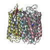
| Title | Crystal structure of the 11-cis isomer of pharaonis halorhodopsin in the absence of halide ions | ||||||
 Components Components | Halorhodopsin | ||||||
 Keywords Keywords | MEMBRANE PROTEIN / seven trans-membrane helices / retinylidene protein / light-driven chloride ion pump microbial rhodopsin | ||||||
| Function / homology | Rhopdopsin 7-helix transmembrane proteins / Rhodopsin 7-helix transmembrane proteins / Up-down Bundle / Mainly Alpha / BACTERIORUBERIN / ISOLEUCINE / Chem-L3P / RETINAL / :  Function and homology information Function and homology information | ||||||
| Biological species |  Natronomonas pharaonis DSM 2160 (archaea) Natronomonas pharaonis DSM 2160 (archaea) | ||||||
| Method |  X-RAY DIFFRACTION / X-RAY DIFFRACTION /  SYNCHROTRON / Resolution: 1.7 Å SYNCHROTRON / Resolution: 1.7 Å | ||||||
 Authors Authors | Kouyama, T. / Chan, S.K. | ||||||
| Funding support |  Japan, 1items Japan, 1items
| ||||||
 Citation Citation |  Journal: Biochemistry / Year: 2016 Journal: Biochemistry / Year: 2016Title: Crystal Structure of the 11-cis Isomer of Pharaonis Halorhodopsin: Structural Constraints on Interconversions among Different Isomeric States Authors: Chan, S.K. / Kawaguchi, H. / Kubo, H. / Murakami, M. / Ihara, K. / Maki, K. / Kouyama, T. | ||||||
| History |
|
- Structure visualization
| Structure viewer | Molecule:  Molmil Molmil Jmol/JSmol Jmol/JSmol |
|---|
- Downloads & links
- Download
| PDBx/mmCIF format |  5b0w.cif.gz 5b0w.cif.gz | 316 KB | Display |  PDBx/mmCIF format PDBx/mmCIF format |
|---|---|---|---|---|
| PDB format |  pdb5b0w.ent.gz pdb5b0w.ent.gz | 253.2 KB | Display |  PDB format PDB format |
| PDBx/mmJSON format |  5b0w.json.gz 5b0w.json.gz | Tree view |  PDBx/mmJSON format PDBx/mmJSON format | |
| Others |  Other downloads Other downloads |
-Validation report
| Summary document |  5b0w_validation.pdf.gz 5b0w_validation.pdf.gz | 3.9 MB | Display |  wwPDB validaton report wwPDB validaton report |
|---|---|---|---|---|
| Full document |  5b0w_full_validation.pdf.gz 5b0w_full_validation.pdf.gz | 4 MB | Display | |
| Data in XML |  5b0w_validation.xml.gz 5b0w_validation.xml.gz | 77.4 KB | Display | |
| Data in CIF |  5b0w_validation.cif.gz 5b0w_validation.cif.gz | 98.5 KB | Display | |
| Arichive directory |  https://data.pdbj.org/pub/pdb/validation_reports/b0/5b0w https://data.pdbj.org/pub/pdb/validation_reports/b0/5b0w ftp://data.pdbj.org/pub/pdb/validation_reports/b0/5b0w ftp://data.pdbj.org/pub/pdb/validation_reports/b0/5b0w | HTTPS FTP |
-Related structure data
- Links
Links
- Assembly
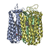
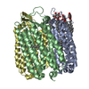
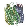
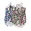
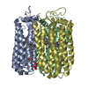
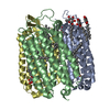
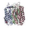
| Deposited unit | 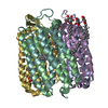
| ||||||||
|---|---|---|---|---|---|---|---|---|---|
| 1 | 
| ||||||||
| 2 | 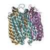
| ||||||||
| Unit cell |
| ||||||||
| Components on special symmetry positions |
|
- Components
-Protein / Sugars , 2 types, 13 molecules ABDEFG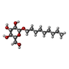

| #1: Protein | Mass: 30975.096 Da / Num. of mol.: 6 / Source method: isolated from a natural source / Source: (natural)  Natronomonas pharaonis DSM 2160 (archaea) / Strain: DSM 2160 / References: UniProt: Q3ITX1 Natronomonas pharaonis DSM 2160 (archaea) / Strain: DSM 2160 / References: UniProt: Q3ITX1#5: Sugar | ChemComp-BNG / |
|---|
-Non-polymers , 5 types, 458 molecules 
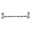
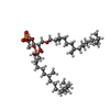


| #2: Chemical | ChemComp-RET / #3: Chemical | ChemComp-22B / | #4: Chemical | ChemComp-L3P / #6: Chemical | ChemComp-ILE / | #7: Water | ChemComp-HOH / | |
|---|
-Details
| Has protein modification | Y |
|---|
-Experimental details
-Experiment
| Experiment | Method:  X-RAY DIFFRACTION / Number of used crystals: 1 X-RAY DIFFRACTION / Number of used crystals: 1 |
|---|
- Sample preparation
| Crystal | Density Matthews: 1.61 Å3/Da / Density % sol: 23.67 % |
|---|---|
| Crystal grow | Temperature: 288 K / Method: vapor diffusion, sitting drop / pH: 6 Details: A mixture solution containing 3mg/ml halorhodopsin, 1M ammonium sulfate, 0.1M NaCl, 0.1 M sodium citrate, and 5.7mg/ml nonylglucoside was concentrated by the sitting-drop vapor diffusion ...Details: A mixture solution containing 3mg/ml halorhodopsin, 1M ammonium sulfate, 0.1M NaCl, 0.1 M sodium citrate, and 5.7mg/ml nonylglucoside was concentrated by the sitting-drop vapor diffusion method. Before data collection, the crystal was soaked in an alkaline solution containing no halide ions, illuminated with red light, and then soaked in a solution at pH 7. |
-Data collection
| Diffraction | Mean temperature: 100 K |
|---|---|
| Diffraction source | Source:  SYNCHROTRON / Site: SYNCHROTRON / Site:  SPring-8 SPring-8  / Beamline: BL38B1 / Wavelength: 1 Å / Beamline: BL38B1 / Wavelength: 1 Å |
| Detector | Type: RAYONIX MX225HE / Detector: CCD / Date: Jun 11, 2015 |
| Radiation | Monochromator: silicon crystal / Protocol: SINGLE WAVELENGTH / Monochromatic (M) / Laue (L): M / Scattering type: x-ray |
| Radiation wavelength | Wavelength: 1 Å / Relative weight: 1 |
| Reflection | Resolution: 1.7→50.7 Å / Num. obs: 125077 / % possible obs: 96.8 % / Observed criterion σ(F): 2.5 / Redundancy: 3.2 % / Biso Wilson estimate: 17.11 Å2 / Rmerge(I) obs: 0.059 / Rsym value: 0.059 / Net I/σ(I): 11.7 |
| Reflection shell | Resolution: 1.7→1.79 Å / Redundancy: 2.4 % / Rmerge(I) obs: 0.393 / Mean I/σ(I) obs: 2.4 / % possible all: 81.8 |
- Processing
| Software |
| ||||||||||||||||||||||||
|---|---|---|---|---|---|---|---|---|---|---|---|---|---|---|---|---|---|---|---|---|---|---|---|---|---|
| Refinement | Resolution: 1.7→15 Å / Cross valid method: FREE R-VALUE / σ(F): 0
| ||||||||||||||||||||||||
| Solvent computation | Bsol: 93.7278 Å2 | ||||||||||||||||||||||||
| Displacement parameters | Biso max: 112.27 Å2 / Biso mean: 22.3454 Å2 / Biso min: 8.34 Å2
| ||||||||||||||||||||||||
| Refinement step | Cycle: final / Resolution: 1.7→15 Å
| ||||||||||||||||||||||||
| Refine LS restraints |
|
 Movie
Movie Controller




 PDBj
PDBj


