 Movie
Movie Controller

[English] 日本語
 Yorodumi
- PDB-4qry: the ground state and the N intermediate of pharaonis halorhodopsi... -
+ Open data

- Basic information
| Entry | Database: PDB / ID: 4qry | ||||||
|---|---|---|---|---|---|---|---|
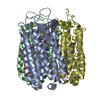
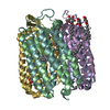
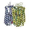
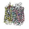
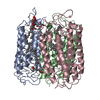
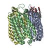
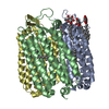
| Title | the ground state and the N intermediate of pharaonis halorhodopsin in complex with bromide ion | ||||||
 Components Components | Halorhodopsin | ||||||
 Keywords Keywords | MEMBRANE PROTEIN / 7 transmembrane helices / light-driven chloride ion pump / retinal bacterioruberin / membrane | ||||||
| Function / homology | Rhopdopsin 7-helix transmembrane proteins / Rhodopsin 7-helix transmembrane proteins / Up-down Bundle / Mainly Alpha / BROMIDE ION / 2,3-DI-PHYTANYL-GLYCEROL / RETINAL / :  Function and homology information Function and homology information | ||||||
| Biological species |  Natronomonas pharaonis (archaea) Natronomonas pharaonis (archaea) | ||||||
| Method |  X-RAY DIFFRACTION / SYNCHROTRON / MOLECULAR REPLACEMENT / Resolution: 2.2 Å X-RAY DIFFRACTION / SYNCHROTRON / MOLECULAR REPLACEMENT / Resolution: 2.2 Å | ||||||
 Authors Authors | Kouyama, T. / Kawaguchi, H. | ||||||
 Citation Citation | Journal: Biophys.J. / Year: 2015 Title: Crystal Structures of the L1, L2, N, and O States of pharaonis Halorhodopsin Authors: Kouyama, T. / Kawaguchi, H. / Nakanishi, T. / Kubo, H. / Murakami, M. | ||||||
| History |
|
- Structure visualization
| Structure viewer | Molecule: MolmilJmol/JSmol |
|---|
- Downloads & links
-Download
| PDBx/mmCIF format | 4qry.cif.gz | 308.7 KB | Display | PDBx/mmCIF format |
|---|---|---|---|---|
| PDB format | pdb4qry.ent.gz | 249.4 KB | Display | PDB format |
| PDBx/mmJSON format | 4qry.json.gz | Tree view | PDBx/mmJSON format | |
| Others |  Other downloads Other downloads |
-Validation report
| Arichive directory | https://data.pdbj.org/pub/pdb/validation_reports/qr/4qryftp://data.pdbj.org/pub/pdb/validation_reports/qr/4qry | HTTPS FTP |
|---|
-Related structure data
| Related structure data | 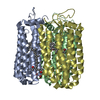 3qbkS S: Starting model for refinement |
|---|---|
| Similar structure data |


-Links
 PDBj
PDBj


- Assembly
| Deposited unit | 
| ||||||||
|---|---|---|---|---|---|---|---|---|---|
| 1 | 
| ||||||||
| 2 | 
| ||||||||
| Unit cell |
|
-Components
-Protein / Sugars , 2 types, 11 molecules ABCEFG
| #1: Protein | Mass: 30975.096 Da / Num. of mol.: 6 / Source method: isolated from a natural source Details: STRAIN MK-1 WAS A HALORHODOPSIN-OVERPRODUCING MUTANT GENERATED FROM TYPE STRAIN D2160T. Source: (natural) Natronomonas pharaonis (archaea) / Strain: MK-1 / References: UniProt: Q3ITX1#4: Sugar | ChemComp-BNG /  Type: D-saccharide / Mass: 306.395 Da / Num. of mol.: 5 Type: D-saccharide / Mass: 306.395 Da / Num. of mol.: 5Source method: isolated from a genetically manipulated source Formula: C15H30O6 / Comment: detergent*YM |
|---|
-Non-polymers , 4 types, 312 molecules 


| #2: Chemical | ChemComp-RET /  Mass: 284.436 Da / Num. of mol.: 6 / Source method: obtained synthetically / Formula: C20H28O Mass: 284.436 Da / Num. of mol.: 6 / Source method: obtained synthetically / Formula: C20H28O#3: Chemical | ChemComp-BR /  Mass: 79.904 Da / Num. of mol.: 12 / Source method: obtained synthetically / Formula: Br Mass: 79.904 Da / Num. of mol.: 12 / Source method: obtained synthetically / Formula: Br#5: Chemical | ChemComp-L2P /  Mass: 653.157 Da / Num. of mol.: 6 / Source method: obtained synthetically / Formula: C43H88O3 Mass: 653.157 Da / Num. of mol.: 6 / Source method: obtained synthetically / Formula: C43H88O3#6: Water | ChemComp-HOH / | Mass: 18.015 Da / Num. of mol.: 288 / Source method: isolated from a natural source / Formula: H2O |
|---|
-Details
| Has protein modification | Y |
|---|
-Experimental details
-Experiment
| Experiment | Method: X-RAY DIFFRACTION / Number of used crystals: 1 |
|---|
- Sample preparation
| Crystal | Density Matthews: 1.6 Å3/Da / Density % sol: 23.3 % |
|---|---|
| Crystal grow | Temperature: 278 K / Method: vapor diffusion, sitting drop / pH: 6 Details: The C2 crystal of halorhodopsin grew when a solution containing 3 mg/ml halorhodopsin, 0.4 % nonylglucoside, 1 M ammonium sulfate, 0.16 M sodium chloride, 0.04 M sodium citrate at pH 6 and 0. ...Details: The C2 crystal of halorhodopsin grew when a solution containing 3 mg/ml halorhodopsin, 0.4 % nonylglucoside, 1 M ammonium sulfate, 0.16 M sodium chloride, 0.04 M sodium citrate at pH 6 and 0.04 % sodium azide was concentrated by the vapor diffusion using a reservoir solution containing 2.8 M ammonium sulfate and 0.12 M sodium citrate at pH 6. For investigation of light-induced structural changes, the C2 crystal was soaked in a solution containing 0.1 M sodium bromide, 1.5 M trisodium citrate at pH 7 and 30 % trehalose. This crystal was illuminated at 240 K with orange light and cooled to 100 K at a rate of 2.3 K/sec in the dark., VAPOR DIFFUSION, SITTING DROP, temperature 278K |
-Data collection
| Diffraction | Mean temperature: 100 K |
|---|---|
| Diffraction source | Source: SYNCHROTRON / Site: SPring-8  / Beamline: BL38B1 / Wavelength: 1 Å / Beamline: BL38B1 / Wavelength: 1 Å |
| Detector | Type: ADSC QUANTUM 315 / Detector: CCD / Date: Jun 3, 2014 |
| Radiation | Monochromator: Si (111) double crystal monochromator / Protocol: SINGLE WAVELENGTH / Monochromatic (M) / Laue (L): M / Scattering type: x-ray |
| Radiation wavelength | Wavelength: 1 Å / Relative weight: 1 |
| Reflection | Resolution: 2.2→49.13 Å / Num. all: 59552 / Num. obs: 58957 / % possible obs: 99 % / Observed criterion σ(F): 2.5 / Observed criterion σ(I): 2.5 / Redundancy: 3.2 % / Biso Wilson estimate: 22.8 Å2 / Rmerge(I) obs: 0.1 / Rsym value: 0.1 / Net I/σ(I): 9.2 |
| Reflection shell | Resolution: 2.2→2.32 Å / Redundancy: 3.3 % / Rmerge(I) obs: 0.5 / Mean I/σ(I) obs: 2.7 / Num. unique all: 8518 / Rsym value: 0.5 / % possible all: 98.5 |
- Processing
| Software |
| |||||||||||||||||||||||||
|---|---|---|---|---|---|---|---|---|---|---|---|---|---|---|---|---|---|---|---|---|---|---|---|---|---|---|
| Refinement | Method to determine structure: MOLECULAR REPLACEMENT Starting model: 3qbk Resolution: 2.2→15 Å / Isotropic thermal model: Isotropic / Cross valid method: THROUGHOUT / σ(F): 0 / Stereochemistry target values: Engh & Huber Details: THE THREE CHAINS (A B AND C) REPRESENT THE REACTION STATES THAT WERE GENERATED BY ILLUMINATION AT 240 K AND REMAINED TRAPPED AFTER SLOW COOLING TO 100K IN THE DARK. THE OTHER THREE CHAINS (E ...Details: THE THREE CHAINS (A B AND C) REPRESENT THE REACTION STATES THAT WERE GENERATED BY ILLUMINATION AT 240 K AND REMAINED TRAPPED AFTER SLOW COOLING TO 100K IN THE DARK. THE OTHER THREE CHAINS (E F AND G) REPRESENT THE UNPHOTOLYZED STATES THAT CO-EXISTED IN THE ILLUMINATED CRYSTAL. THE ASYMMETRIC UNIT OF THE C2 CRYSTAL CONTAINS THREE SUBUNITS. THESE THREE SUBUNITS UNDERWENT DIFFERENT STRUCTURAL CHANGES WHEN THE CRYSTAL WAS ILLUMINATED AT 240K. THE C2 CRYSTAL HAD BEEN SOAKED IN 0.1M SODIUM BROMIDE, 1.5M TRISODIUM-CITRATE AT pH 7 AND 30% TREHALOSE. THE DIFFRACTION DATA WERE COLLECTED AFTER THE ILLUMINATED CRYSTAL WAS COOLED TO 100 K WITH A RATE 2.3K/SEC IN THE DARK.
| |||||||||||||||||||||||||
| Displacement parameters | Biso mean: 24.9699 Å2
| |||||||||||||||||||||||||
| Refine analyze |
| |||||||||||||||||||||||||
| Refinement step | Cycle: LAST / Resolution: 2.2→15 Å
| |||||||||||||||||||||||||
| Refine LS restraints |
|
