 Movie
Movie Controller

[English] 日本語
 Yorodumi
- PDB-3abw: Crystal structure of pharaonis halorhodopsin in complex with azide ion -
+ Open data

- Basic information
| Entry | Database: PDB / ID: 3abw | ||||||
|---|---|---|---|---|---|---|---|
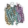
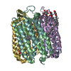
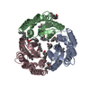
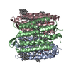
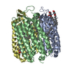
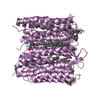
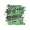
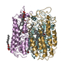
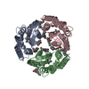
| Title | Crystal structure of pharaonis halorhodopsin in complex with azide ion | ||||||
 Components Components | Halorhodopsin | ||||||
 Keywords Keywords | MEMBRANE PROTEIN / LIGHT-DRIVEN CHLORIDE ION PUMP / TRIMERIC BACTERIORUBERIN-PROTEIN COMPLEX / RETINAL PROTEIN | ||||||
| Function / homology | Rhopdopsin 7-helix transmembrane proteins / Rhodopsin 7-helix transmembrane proteins / Up-down Bundle / Mainly Alpha / BACTERIORUBERIN / AZIDE ION / Chem-L3P / RETINAL / :  Function and homology information Function and homology information | ||||||
| Biological species |  Natronomonas pharaonis (archaea) Natronomonas pharaonis (archaea) | ||||||
| Method |  X-RAY DIFFRACTION / SYNCHROTRON / MOLECULAR REPLACEMENT / Resolution: 1.9 Å X-RAY DIFFRACTION / SYNCHROTRON / MOLECULAR REPLACEMENT / Resolution: 1.9 Å | ||||||
 Authors Authors | Kouyama, T. / Kanada, S. | ||||||
 Citation Citation | Journal: To be Published Title: Binding mode of azide ion to pharaonis halorhodopsin Authors: Kanada, S. / Kouyama, T. | ||||||
| History |
|
- Structure visualization
| Structure viewer | Molecule: MolmilJmol/JSmol |
|---|
- Downloads & links
-Download
| PDBx/mmCIF format | 3abw.cif.gz | 169.5 KB | Display | PDBx/mmCIF format |
|---|---|---|---|---|
| PDB format | pdb3abw.ent.gz | 131.1 KB | Display | PDB format |
| PDBx/mmJSON format | 3abw.json.gz | Tree view | PDBx/mmJSON format | |
| Others |  Other downloads Other downloads |
-Validation report
| Arichive directory | https://data.pdbj.org/pub/pdb/validation_reports/ab/3abwftp://data.pdbj.org/pub/pdb/validation_reports/ab/3abw | HTTPS FTP |
|---|
-Related structure data
| Related structure data |  3a7kS S: Starting model for refinement |
|---|---|
| Similar structure data |

-Links
 PDBj
PDBj


- Assembly
| Deposited unit | 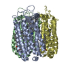
| ||||||||
|---|---|---|---|---|---|---|---|---|---|
| 1 |
| ||||||||
| Unit cell |
|
-Components
-Protein , 1 types, 3 molecules ABD
| #1: Protein | Mass: 30975.096 Da / Num. of mol.: 3 / Source method: isolated from a natural source Details: STRAIN MK-1 WAS A HALORHODOPSIN-OVERPRODUCING MUTANT GENERATED FROM TYPE STRAIN D2160T. Source: (natural) Natronomonas pharaonis (archaea) / Strain: MK-1 / References: UniProt: Q3ITX1 |
|---|
-Non-polymers , 5 types, 145 molecules 


| #2: Chemical |  Mass: 284.436 Da / Num. of mol.: 3 / Source method: obtained synthetically / Formula: C20H28O Mass: 284.436 Da / Num. of mol.: 3 / Source method: obtained synthetically / Formula: C20H28O#3: Chemical |  Mass: 741.136 Da / Num. of mol.: 2 / Source method: obtained synthetically / Formula: C50H76O4 Mass: 741.136 Da / Num. of mol.: 2 / Source method: obtained synthetically / Formula: C50H76O4#4: Chemical | ChemComp-L3P /  Mass: 885.179 Da / Num. of mol.: 9 / Source method: obtained synthetically / Formula: C46H94O11P2 Mass: 885.179 Da / Num. of mol.: 9 / Source method: obtained synthetically / Formula: C46H94O11P2#5: Chemical |  Mass: 42.020 Da / Num. of mol.: 3 / Source method: obtained synthetically / Formula: N3 Mass: 42.020 Da / Num. of mol.: 3 / Source method: obtained synthetically / Formula: N3#6: Water | ChemComp-HOH / | Mass: 18.015 Da / Num. of mol.: 128 / Source method: isolated from a natural source / Formula: H2O |
|---|
-Details
| Has protein modification | Y |
|---|
-Experimental details
-Experiment
| Experiment | Method: X-RAY DIFFRACTION / Number of used crystals: 1 |
|---|
- Sample preparation
| Crystal | Density Matthews: 3.2 Å3/Da / Density % sol: 61.52 % |
|---|---|
| Crystal grow | Temperature: 288 K / Method: vapor diffusion, sitting drop / pH: 6 Details: A DROP CONTAINING 2MG/ML Halorhodopsin, 1M AMMONIUM SULFATE, 0.16M NACL, 0.04M Na-citrate, 0.04% NAN3,5MG/ML NONYL GLUCOSIDE WAS CONCENTRATED BY VAPOR DIFFUSIONAGAINST A RESERVE SOLUTION ...Details: A DROP CONTAINING 2MG/ML Halorhodopsin, 1M AMMONIUM SULFATE, 0.16M NACL, 0.04M Na-citrate, 0.04% NAN3,5MG/ML NONYL GLUCOSIDE WAS CONCENTRATED BY VAPOR DIFFUSIONAGAINST A RESERVE SOLUTION CONTAINING 2.9 M AMMONIUM SULFATE, 0.04M Na-citrate, PH 6, VAPOR DIFFUSION, SITTING DROP, TEMPERATURE 288K. After the crystal grew, the mother solution was exchanged with a solution containing 3M Ammonium sulfate, 200 mM Na-N3, 100 mM HEPES (pH7) and 30 % trehalose. |
-Data collection
| Diffraction | Mean temperature: 100 K |
|---|---|
| Diffraction source | Source: SYNCHROTRON / Site: SPring-8  / Beamline: BL38B1 / Wavelength: 1 Å / Beamline: BL38B1 / Wavelength: 1 Å |
| Detector | Type: RIGAKU JUPITER 210 / Detector: CCD / Date: Dec 14, 2009 / Details: mirrors |
| Radiation | Monochromator: Si 111 / Protocol: SINGLE WAVELENGTH / Monochromatic (M) / Laue (L): M / Scattering type: x-ray |
| Radiation wavelength | Wavelength: 1 Å / Relative weight: 1 |
| Reflection | Resolution: 1.9→38.15 Å / Num. all: 91795 / Num. obs: 80799 / % possible obs: 88 % / Observed criterion σ(F): 0 / Observed criterion σ(I): 0 / Redundancy: 3.2 % / Biso Wilson estimate: 23.3 Å2 / Rmerge(I) obs: 0.071 / Rsym value: 0.071 / Net I/σ(I): 10.5 |
| Reflection shell | Resolution: 1.9→2 Å / Redundancy: 3 % / Rmerge(I) obs: 0.526 / Mean I/σ(I) obs: 2.2 / Rsym value: 0.526 / % possible all: 74.3 |
- Processing
| Software |
| |||||||||||||||||||||||||||||||||
|---|---|---|---|---|---|---|---|---|---|---|---|---|---|---|---|---|---|---|---|---|---|---|---|---|---|---|---|---|---|---|---|---|---|---|
| Refinement | Method to determine structure: MOLECULAR REPLACEMENT Starting model: PDB ENTRY 3a7k Resolution: 1.9→15 Å / Cross valid method: THROUGHOUT / σ(F): 0 / σ(I): 0 / Stereochemistry target values: Engh & Huber
| |||||||||||||||||||||||||||||||||
| Solvent computation | Bsol: 99.0968 Å2 | |||||||||||||||||||||||||||||||||
| Displacement parameters | Biso mean: 32.4952 Å2
| |||||||||||||||||||||||||||||||||
| Refine analyze |
| |||||||||||||||||||||||||||||||||
| Refinement step | Cycle: LAST / Resolution: 1.9→15 Å
| |||||||||||||||||||||||||||||||||
| Refine LS restraints |
| |||||||||||||||||||||||||||||||||
| LS refinement shell |
| |||||||||||||||||||||||||||||||||
| Xplor file |
|
