 Movie
Movie Controller

[English] 日本語
 Yorodumi
- PDB-5kkh: 2.1-Angstrom In situ Mylar structure of bacteriorhodopsin from Ha... -
+ Open data

- Basic information
| Entry | Database: PDB / ID: 5kkh | |||||||||
|---|---|---|---|---|---|---|---|---|---|---|
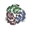
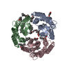
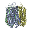
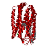
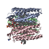
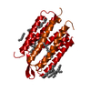
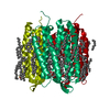
| Title | 2.1-Angstrom In situ Mylar structure of bacteriorhodopsin from Haloquadratum walsbyi (HwBR) at 100 K | |||||||||
 Components Components | Bacteriorhodopsin-I | |||||||||
 Keywords Keywords | MEMBRANE PROTEIN / TRANSPORT PROTEIN | |||||||||
| Function / homology |  Function and homology information Function and homology informationphotoreceptor activity / phototransduction / monoatomic ion channel activity / proton transmembrane transport / plasma membrane Similarity search - Function | |||||||||
| Biological species |  Haloquadratum walsbyi (archaea) Haloquadratum walsbyi (archaea) | |||||||||
| Method |  X-RAY DIFFRACTION / SYNCHROTRON / MOLECULAR REPLACEMENT / Resolution: 2.125 Å X-RAY DIFFRACTION / SYNCHROTRON / MOLECULAR REPLACEMENT / Resolution: 2.125 Å | |||||||||
 Authors Authors | Broecker, J. / Ernst, O.P. | |||||||||
| Funding support |  Canada, Canada,  Germany, 2items Germany, 2items
| |||||||||
 Citation Citation | Journal: Cryst Growth Des / Year: 2016 Title: A Versatile System for High-Throughput In Situ X-ray Screening and Data Collection of Soluble and Membrane-Protein Crystals. Authors: Broecker, J. / Klingel, V. / Ou, W.L. / Balo, A.R. / Kissick, D.J. / Ogata, C.M. / Kuo, A. / Ernst, O.P. | |||||||||
| History |
|
- Structure visualization
| Structure viewer | Molecule: MolmilJmol/JSmol |
|---|
- Downloads & links
-Download
| PDBx/mmCIF format | 5kkh.cif.gz | 151.5 KB | Display | PDBx/mmCIF format |
|---|---|---|---|---|
| PDB format | pdb5kkh.ent.gz | 120.7 KB | Display | PDB format |
| PDBx/mmJSON format | 5kkh.json.gz | Tree view | PDBx/mmJSON format | |
| Others |  Other downloads Other downloads |
-Validation report
| Arichive directory | https://data.pdbj.org/pub/pdb/validation_reports/kk/5kkhftp://data.pdbj.org/pub/pdb/validation_reports/kk/5kkh | HTTPS FTP |
|---|
-Related structure data
| Related structure data |  5kkiC  5kkjC 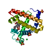 5kkkC  2ei4S C: citing same article ( S: Starting model for refinement |
|---|---|
| Similar structure data |


-Links
 PDBj
PDBj


- Assembly
| Deposited unit | 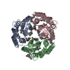
| ||||||||
|---|---|---|---|---|---|---|---|---|---|
| 1 |
| ||||||||
| Unit cell |
|
-Components
| #1: Protein | Mass: 29116.809 Da / Num. of mol.: 3 Source method: isolated from a genetically manipulated source Source: (gene. exp.) Haloquadratum walsbyi (strain DSM 16790 / HBSQ001) (archaea)Strain: DSM 16790 / HBSQ001 / Gene: bop1, bopI, HQ_1014A / Production host:  #2: Chemical | ChemComp-OLC / (   Mass: 356.540 Da / Num. of mol.: 6 / Source method: obtained synthetically / Formula: C21H40O4 Mass: 356.540 Da / Num. of mol.: 6 / Source method: obtained synthetically / Formula: C21H40O4#3: Chemical |   Mass: 356.540 Da / Num. of mol.: 3 / Source method: obtained synthetically / Formula: C21H40O4 Mass: 356.540 Da / Num. of mol.: 3 / Source method: obtained synthetically / Formula: C21H40O4#4: Chemical |   Mass: 284.436 Da / Num. of mol.: 3 / Source method: obtained synthetically / Formula: C20H28O Mass: 284.436 Da / Num. of mol.: 3 / Source method: obtained synthetically / Formula: C20H28O#5: Water | ChemComp-HOH / |  Mass: 18.015 Da / Num. of mol.: 84 / Source method: isolated from a natural source / Formula: H2O Mass: 18.015 Da / Num. of mol.: 84 / Source method: isolated from a natural source / Formula: H2OHas protein modification | Y | |
|---|
-Experimental details
-Experiment
| Experiment | Method: X-RAY DIFFRACTION / Number of used crystals: 1 |
|---|
- Sample preparation
| Crystal | Density Matthews: 1.97 Å3/Da / Density % sol: 37.7 % / Description: small compact, hexagonal plates |
|---|---|
| Crystal grow | Temperature: 293 K / Method: lipidic cubic phase / pH: 7 / Details: 8% (v/v) Tacsimate, 20% (v/v) PEG 3350 |
-Data collection
| Diffraction | Mean temperature: 100 K |
|---|---|
| Diffraction source | Source: SYNCHROTRON / Site: APS  / Beamline: 23-ID-D / Wavelength: 1 Å / Beamline: 23-ID-D / Wavelength: 1 Å |
| Detector | Type: DECTRIS PILATUS 6M / Detector: PIXEL / Date: Mar 11, 2016 |
| Radiation | Protocol: SINGLE WAVELENGTH / Monochromatic (M) / Laue (L): M / Scattering type: x-ray |
| Radiation wavelength | Wavelength: 1 Å / Relative weight: 1 |
| Reflection | Resolution: 2.125→29.45 Å / Num. all: 57569 / Num. obs: 35323 / % possible obs: 91 % / Redundancy: 1.8 % / Biso Wilson estimate: 14.86 Å2 / CC1/2: 0.96 / Rmerge(I) obs: 0.107 / Net I/σ(I): 6.26 |
| Reflection shell | Resolution: 2.13→2.2 Å / Redundancy: 1.8 % / Rmerge(I) obs: 0.25 / Mean I/σ(I) obs: 3.66 / % possible all: 70 |
- Processing
| Software |
| ||||||||||||||||||||||||||||||||||||||||||||||||||||||||||||||||||||||||||||||||||||||||||||||||||
|---|---|---|---|---|---|---|---|---|---|---|---|---|---|---|---|---|---|---|---|---|---|---|---|---|---|---|---|---|---|---|---|---|---|---|---|---|---|---|---|---|---|---|---|---|---|---|---|---|---|---|---|---|---|---|---|---|---|---|---|---|---|---|---|---|---|---|---|---|---|---|---|---|---|---|---|---|---|---|---|---|---|---|---|---|---|---|---|---|---|---|---|---|---|---|---|---|---|---|---|
| Refinement | Method to determine structure: MOLECULAR REPLACEMENT Starting model: 2EI4 Resolution: 2.125→29.446 Å / SU ML: 0.18 / Cross valid method: FREE R-VALUE / σ(F): 1.34 / Phase error: 20.95 / Stereochemistry target values: ML
| ||||||||||||||||||||||||||||||||||||||||||||||||||||||||||||||||||||||||||||||||||||||||||||||||||
| Solvent computation | Shrinkage radii: 0.9 Å / VDW probe radii: 1.11 Å / Solvent model: FLAT BULK SOLVENT MODEL | ||||||||||||||||||||||||||||||||||||||||||||||||||||||||||||||||||||||||||||||||||||||||||||||||||
| Refinement step | Cycle: LAST / Resolution: 2.125→29.446 Å
| ||||||||||||||||||||||||||||||||||||||||||||||||||||||||||||||||||||||||||||||||||||||||||||||||||
| Refine LS restraints |
| ||||||||||||||||||||||||||||||||||||||||||||||||||||||||||||||||||||||||||||||||||||||||||||||||||
| LS refinement shell |
|
