 Movie
Movie Controller

[English] 日本語
 Yorodumi
- PDB-6lm0: The crystal structure of cyanorhodopsin (CyR) N2098R from cyanoba... -
+ Open data

- Basic information
| Entry | Database: PDB / ID: 6lm0 | ||||||
|---|---|---|---|---|---|---|---|
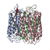
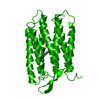
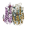
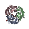
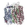
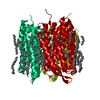
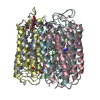
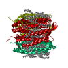
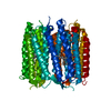
| Title | The crystal structure of cyanorhodopsin (CyR) N2098R from cyanobacteria Calothrix sp. NIES-2098 | ||||||
 Components Components | Rhodopsin | ||||||
 Keywords Keywords | MEMBRANE PROTEIN / RETINAL CELL-FREE SYNTHESIS Bacterial type rhodopsin Cyanobacteria | ||||||
| Function / homology |  Function and homology information Function and homology information: / photoreceptor activity / phototransduction / membrane => GO:0016020 / monoatomic ion channel activity Similarity search - Function | ||||||
| Biological species |  Calothrix sp. NIES-2098 (bacteria) Calothrix sp. NIES-2098 (bacteria) | ||||||
| Method |  X-RAY DIFFRACTION / SYNCHROTRON / MOLECULAR REPLACEMENT / Resolution: 2.65 Å X-RAY DIFFRACTION / SYNCHROTRON / MOLECULAR REPLACEMENT / Resolution: 2.65 Å | ||||||
 Authors Authors | Hosaka, T. / Kimura-Someya, T. / Shirouzu, M. | ||||||
 Citation Citation | Journal: Sci Rep / Year: 2020 Title: A unique clade of light-driven proton-pumping rhodopsins evolved in the cyanobacterial lineage. Authors: Hasegawa, M. / Hosaka, T. / Kojima, K. / Nishimura, Y. / Nakajima, Y. / Kimura-Someya, T. / Shirouzu, M. / Sudo, Y. / Yoshizawa, S. | ||||||
| History |
|
- Structure visualization
| Structure viewer | Molecule: MolmilJmol/JSmol |
|---|
- Downloads & links
-Download
| PDBx/mmCIF format | 6lm0.cif.gz | 155 KB | Display | PDBx/mmCIF format |
|---|---|---|---|---|
| PDB format | pdb6lm0.ent.gz | 120.9 KB | Display | PDB format |
| PDBx/mmJSON format | 6lm0.json.gz | Tree view | PDBx/mmJSON format | |
| Others |  Other downloads Other downloads |
-Validation report
| Arichive directory | https://data.pdbj.org/pub/pdb/validation_reports/lm/6lm0ftp://data.pdbj.org/pub/pdb/validation_reports/lm/6lm0 | HTTPS FTP |
|---|
-Related structure data
| Related structure data | 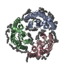 6lm1C 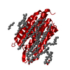 1c3wS S: Starting model for refinement C: citing same article ( |
|---|---|
| Similar structure data |

-Links
 PDBj
PDBj


- Assembly
| Deposited unit | 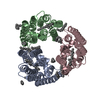
| ||||||||
|---|---|---|---|---|---|---|---|---|---|
| 1 |
| ||||||||
| Unit cell |
|
-Components
-Protein , 1 types, 3 molecules ABC
| #1: Protein | Mass: 28253.928 Da / Num. of mol.: 3 Source method: isolated from a genetically manipulated source Source: (gene. exp.) Calothrix sp. NIES-2098 (bacteria) / Gene: NIES2098_21630 / Production host: |
|---|
-Non-polymers , 6 types, 75 molecules 


| #2: Chemical |  Mass: 284.436 Da / Num. of mol.: 3 / Source method: obtained synthetically / Formula: C20H28O / Feature type: SUBJECT OF INVESTIGATION Mass: 284.436 Da / Num. of mol.: 3 / Source method: obtained synthetically / Formula: C20H28O / Feature type: SUBJECT OF INVESTIGATION#3: Chemical |  Mass: 86.175 Da / Num. of mol.: 3 / Source method: obtained synthetically / Formula: C6H14 / Feature type: SUBJECT OF INVESTIGATION Mass: 86.175 Da / Num. of mol.: 3 / Source method: obtained synthetically / Formula: C6H14 / Feature type: SUBJECT OF INVESTIGATION#4: Chemical | ChemComp-OCT /  Mass: 114.229 Da / Num. of mol.: 4 / Source method: obtained synthetically / Formula: C8H18 / Feature type: SUBJECT OF INVESTIGATION Mass: 114.229 Da / Num. of mol.: 4 / Source method: obtained synthetically / Formula: C8H18 / Feature type: SUBJECT OF INVESTIGATION#5: Chemical | ChemComp-C14 /  Mass: 198.388 Da / Num. of mol.: 4 / Source method: obtained synthetically / Formula: C14H30 / Feature type: SUBJECT OF INVESTIGATION Mass: 198.388 Da / Num. of mol.: 4 / Source method: obtained synthetically / Formula: C14H30 / Feature type: SUBJECT OF INVESTIGATION#6: Chemical |  Mass: 142.282 Da / Num. of mol.: 2 / Source method: obtained synthetically / Formula: C10H22 / Feature type: SUBJECT OF INVESTIGATION Mass: 142.282 Da / Num. of mol.: 2 / Source method: obtained synthetically / Formula: C10H22 / Feature type: SUBJECT OF INVESTIGATION#7: Water | ChemComp-HOH / | Mass: 18.015 Da / Num. of mol.: 59 / Source method: isolated from a natural source / Formula: H2O |
|---|
-Details
| Has ligand of interest | Y |
|---|---|
| Has protein modification | Y |
-Experimental details
-Experiment
| Experiment | Method: X-RAY DIFFRACTION / Number of used crystals: 1 |
|---|
- Sample preparation
| Crystal | Density Matthews: 2.3 Å3/Da / Density % sol: 46.45 % |
|---|---|
| Crystal grow | Temperature: 293 K / Method: lipidic cubic phase Details: 0.1M sodium acetate (pH5.0), 200mM sodiumu chloride, 39% PEG 500 |
-Data collection
| Diffraction | Mean temperature: 100 K / Serial crystal experiment: N | ||||||||||||||||||||||||||||||||||||||||||||||||||||||||||||||||||||||||||||||||||||||||||||||||||||
|---|---|---|---|---|---|---|---|---|---|---|---|---|---|---|---|---|---|---|---|---|---|---|---|---|---|---|---|---|---|---|---|---|---|---|---|---|---|---|---|---|---|---|---|---|---|---|---|---|---|---|---|---|---|---|---|---|---|---|---|---|---|---|---|---|---|---|---|---|---|---|---|---|---|---|---|---|---|---|---|---|---|---|---|---|---|---|---|---|---|---|---|---|---|---|---|---|---|---|---|---|---|
| Diffraction source | Source: SYNCHROTRON / Site: SPring-8  / Beamline: BL32XU / Wavelength: 1 Å / Beamline: BL32XU / Wavelength: 1 Å | ||||||||||||||||||||||||||||||||||||||||||||||||||||||||||||||||||||||||||||||||||||||||||||||||||||
| Detector | Type: DECTRIS EIGER X 9M / Detector: PIXEL / Date: Feb 1, 2019 | ||||||||||||||||||||||||||||||||||||||||||||||||||||||||||||||||||||||||||||||||||||||||||||||||||||
| Radiation | Protocol: SINGLE WAVELENGTH / Monochromatic (M) / Laue (L): M / Scattering type: x-ray | ||||||||||||||||||||||||||||||||||||||||||||||||||||||||||||||||||||||||||||||||||||||||||||||||||||
| Radiation wavelength | Wavelength: 1 Å / Relative weight: 1 | ||||||||||||||||||||||||||||||||||||||||||||||||||||||||||||||||||||||||||||||||||||||||||||||||||||
| Reflection | Resolution: 2.65→49.615 Å / Num. obs: 23218 / % possible obs: 100 % / Redundancy: 72.416 % / Biso Wilson estimate: 45.065 Å2 / CC1/2: 0.993 / Rmerge(I) obs: 0.727 / Rrim(I) all: 0.732 / Χ2: 1.222 / Net I/σ(I): 10.76 / Num. measured all: 1681366 / Scaling rejects: 446 | ||||||||||||||||||||||||||||||||||||||||||||||||||||||||||||||||||||||||||||||||||||||||||||||||||||
| Reflection shell | Diffraction-ID: 1
|
- Processing
| Software |
| ||||||||||||||||||||||||||||||||||||||||||||||||||||||||||||||||||||||||||||||||||||||||||
|---|---|---|---|---|---|---|---|---|---|---|---|---|---|---|---|---|---|---|---|---|---|---|---|---|---|---|---|---|---|---|---|---|---|---|---|---|---|---|---|---|---|---|---|---|---|---|---|---|---|---|---|---|---|---|---|---|---|---|---|---|---|---|---|---|---|---|---|---|---|---|---|---|---|---|---|---|---|---|---|---|---|---|---|---|---|---|---|---|---|---|---|
| Refinement | Method to determine structure: MOLECULAR REPLACEMENT Starting model: 1C3W Resolution: 2.65→49.615 Å / SU ML: 0.33 / Cross valid method: THROUGHOUT / σ(F): 1.33 / Phase error: 26.63 / Stereochemistry target values: ML
| ||||||||||||||||||||||||||||||||||||||||||||||||||||||||||||||||||||||||||||||||||||||||||
| Solvent computation | Shrinkage radii: 0.9 Å / VDW probe radii: 1.11 Å / Solvent model: FLAT BULK SOLVENT MODEL | ||||||||||||||||||||||||||||||||||||||||||||||||||||||||||||||||||||||||||||||||||||||||||
| Displacement parameters | Biso max: 98.95 Å2 / Biso mean: 41.2469 Å2 / Biso min: 17.71 Å2 | ||||||||||||||||||||||||||||||||||||||||||||||||||||||||||||||||||||||||||||||||||||||||||
| Refinement step | Cycle: final / Resolution: 2.65→49.615 Å
| ||||||||||||||||||||||||||||||||||||||||||||||||||||||||||||||||||||||||||||||||||||||||||
| LS refinement shell | Refine-ID: X-RAY DIFFRACTION / Rfactor Rfree error: 0
|
