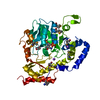
PDB-3tq8: Structure of the dihydrofolate reductase (folA) from Coxiella burnetii in complex with trimethoprim 手法: X-RAY DIFFRACTION / 解像度: 1.9 Å
PDB-3tq9: Structure of the dihydrofolate reductase (folA) from Coxiella burnetii in complex with methotrexate 手法: X-RAY DIFFRACTION / 解像度: 2.3 Å
PDB-3tqa: Structure of the dihydrofolate reductase (folA) from Coxiella burnetii in complex with NADPH 手法: X-RAY DIFFRACTION / 解像度: 2.3 Å
PDB-3tqb: Structure of the dihydrofolate reductase (folA) from Coxiella burnetii in complex with folate 手法: X-RAY DIFFRACTION / 解像度: 2.4 Å
PDB-3tqc: Structure of the pantothenate kinase (coaA) from Coxiella burnetii 手法: X-RAY DIFFRACTION / 解像度: 2.3 Å
PDB-3tqd: Structure of the 3-deoxy-D-manno-octulosonate cytidylyltransferase (kdsB) from Coxiella burnetii 手法: X-RAY DIFFRACTION / 解像度: 1.8 Å
PDB-3tqe: Structure of the malonyl CoA-acyl carrier protein transacylase (fabD) from Coxiella burnetii 手法: X-RAY DIFFRACTION / 解像度: 1.5 Å
PDB-3tqf: Structure of the Hpr(Ser) kinase/phosphatase from Coxiella burnetii 手法: X-RAY DIFFRACTION / 解像度: 2.8 Å
PDB-3tqg: Structure of the 2-methylcitrate synthase (prpC) from Coxiella burnetii 手法: X-RAY DIFFRACTION / 解像度: 2.3 Å
PDB-3tqh: Structure of the quinone oxidoreductase from Coxiella burnetii 手法: X-RAY DIFFRACTION / 解像度: 2.44 Å
PDB-3tqi: Structure of the GMP synthase (guaA) from Coxiella burnetii 手法: X-RAY DIFFRACTION / 解像度: 2.84 Å
PDB-3tqj: Structure of the superoxide dismutase (Fe) (sodB) from Coxiella burnetii 手法: X-RAY DIFFRACTION / 解像度: 2.004 Å
PDB-3tql: Structure of the amino acid ABC transporter, periplasmic amino acid-binding protein from Coxiella burnetii. 手法: X-RAY DIFFRACTION / 解像度: 1.594 Å
PDB-3tqm: Structure of an ribosomal subunit interface protein from Coxiella burnetii 手法: X-RAY DIFFRACTION / 解像度: 2.45 Å
PDB-3tqn: Structure of the transcriptional regulator of the GntR family, from Coxiella burnetii. 手法: X-RAY DIFFRACTION / 解像度: 2.8 Å
PDB-3tqo: Structure of the cysteinyl-tRNA synthetase (cysS) from Coxiella burnetii. 手法: X-RAY DIFFRACTION / 解像度: 2.3 Å
PDB-3tqp: Structure of an enolase (eno) from Coxiella burnetii 手法: X-RAY DIFFRACTION / 解像度: 2.2 Å
PDB-3tqq: Structure of the methionyl-tRNA formyltransferase (fmt) from Coxiella burnetii 手法: X-RAY DIFFRACTION / 解像度: 2 Å
PDB-3tqr: Structure of the phosphoribosylglycinamide formyltransferase (purN) in complex with CHES from Coxiella burnetii 手法: X-RAY DIFFRACTION / 解像度: 1.97 Å
PDB-3tqs: Structure of the dimethyladenosine transferase (ksgA) from Coxiella burnetii 手法: X-RAY DIFFRACTION / 解像度: 1.98 Å
PDB-3tqt: Structure of the D-alanine-D-alanine ligase from Coxiella burnetii 手法: X-RAY DIFFRACTION / 解像度: 1.88 Å
PDB-3tqu: Structure of a HAM1 protein from Coxiella burnetii 手法: X-RAY DIFFRACTION / 解像度: 1.9 Å
PDB-3tqw: Structure of a ABC transporter, periplasmic substrate-binding protein from Coxiella burnetii 手法: X-RAY DIFFRACTION / 解像度: 2 Å
PDB-3tqx: Structure of the 2-amino-3-ketobutyrate coenzyme A ligase (kbl) from Coxiella burnetii 手法: X-RAY DIFFRACTION / 解像度: 2.304 Å
PDB-3tqy: Structure of a single-stranded DNA-binding protein (ssb), from Coxiella burnetii 手法: X-RAY DIFFRACTION / 解像度: 2.6001 Å
PDB-3tqz: Structure of a deoxyuridine 5'-triphosphate nucleotidohydrolase (dut) from Coxiella burnetii 手法: X-RAY DIFFRACTION / 解像度: 1.75 Å
PDB-3tr0: Structure of guanylate kinase (gmk) from Coxiella burnetii 手法: X-RAY DIFFRACTION / 解像度: 1.851 Å
PDB-3tr1: Structure of a 3-phosphoshikimate 1-carboxyvinyltransferase (aroA) from Coxiella burnetii 手法: X-RAY DIFFRACTION / 解像度: 2 Å
PDB-3tr2: Structure of a orotidine 5'-phosphate decarboxylase (pyrF) from Coxiella burnetii 手法: X-RAY DIFFRACTION / 解像度: 2.001 Å
PDB-3tr3: Structure of a bolA protein homologue from Coxiella burnetii 手法: X-RAY DIFFRACTION / 解像度: 2.456 Å
PDB-3tr4: Structure of an inorganic pyrophosphatase (ppa) from Coxiella burnetii 手法: X-RAY DIFFRACTION / 解像度: 2 Å
PDB-3tr5: Structure of a peptide chain release factor 3 (prfC) from Coxiella burnetii 手法: X-RAY DIFFRACTION / 解像度: 2.11 Å
PDB-3tr6: Structure of a O-methyltransferase from Coxiella burnetii 手法: X-RAY DIFFRACTION / 解像度: 2.7 Å
PDB-3tr7: Structure of a uracil-DNA glycosylase (ung) from Coxiella burnetii 手法: X-RAY DIFFRACTION / 解像度: 2.1958 Å
PDB-3tr8: Structure of an oligoribonuclease (orn) from Coxiella burnetii 手法: X-RAY DIFFRACTION / 解像度: 2.503 Å
PDB-3tr9: Structure of a dihydropteroate synthase (folP) in complex with pteroic acid from Coxiella burnetii 手法: X-RAY DIFFRACTION / 解像度: 1.895 Å
PDB-3trb: Structure of an addiction module antidote protein of a HigA (higA) family from Coxiella burnetii 手法: X-RAY DIFFRACTION / 解像度: 2.001 Å
PDB-3trc: Structure of the GAF domain from a phosphoenolpyruvate-protein phosphotransferase (ptsP) from Coxiella burnetii 手法: X-RAY DIFFRACTION / 解像度: 1.65 Å
PDB-3trd: Structure of an alpha-beta serine hydrolase homologue from Coxiella burnetii 手法: X-RAY DIFFRACTION / 解像度: 1.5 Å
PDB-3tre: Structure of a translation elongation factor P (efp) from Coxiella burnetii 手法: X-RAY DIFFRACTION / 解像度: 2.899 Å
PDB-3trf: Structure of a shikimate kinase (aroK) from Coxiella burnetii 手法: X-RAY DIFFRACTION / 解像度: 2.6 Å
PDB-3trg: Structure of an acylphosphatase from Coxiella burnetii 手法: X-RAY DIFFRACTION / 解像度: 1.601 Å
PDB-3trh: Structure of a phosphoribosylaminoimidazole carboxylase catalytic subunit (purE) from Coxiella burnetii 手法: X-RAY DIFFRACTION / 解像度: 2.203 Å
PDB-3tri: Structure of a pyrroline-5-carboxylate reductase (proC) from Coxiella burnetii 手法: X-RAY DIFFRACTION / 解像度: 2.5 Å
PDB-3tth: Structure of the spermidine N1-acetyltransferase (speG) from Coxiella burnetii 手法: X-RAY DIFFRACTION / 解像度: 3.298 Å
PDB-3ty2: Structure of a 5'-nucleotidase (surE) from Coxiella burnetii 手法: X-RAY DIFFRACTION / 解像度: 1.885 Å
PDB-3uwc: Structure of an aminotransferase (DegT-DnrJ-EryC1-StrS family) from Coxiella burnetii in complex with PMP 手法: X-RAY DIFFRACTION / 解像度: 1.8 Å
PDB-4f3q: Structure of a YebC family protein (CBU_1566) from Coxiella burnetii 手法: X-RAY DIFFRACTION / 解像度: 2.15 Å
PDB-4f3r: Structure of phosphopantetheine adenylyltransferase (CBU_0288) from Coxiella burnetii 手法: X-RAY DIFFRACTION / 解像度: 2.25 Å
PDB-4nbq: Structure of the polynucleotide phosphorylase (CBU_0852) from Coxiella burnetii 手法: X-RAY DIFFRACTION / 解像度: 2.9138 Å
PDB-4ng4: Structure of phosphoglycerate kinase (CBU_1782) from Coxiella burnetii 手法: X-RAY DIFFRACTION / 解像度: 2.78 Å
 ムービー
ムービー コントローラー
 構造ビューア
 万見文献について


 著者
著者 リンク
リンク


 キーワード
キーワード coxiella burnetii (バクテリア)