 ムービー
ムービー コントローラー
 構造ビューア
 EMN検索について


-検索条件
-検索結果
検索 (著者・登録者: sieber & s)の結果102件中、1から50件目までを表示しています

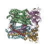
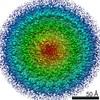
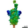
EMDB-15616: 
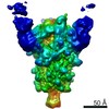
Four subunit cytochrome b-c1 complex from Rhodobacter sphaeroides in native nanodiscs - consensus refinement in the b-b conformation

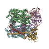
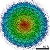
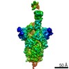
EMDB-15617:
Four subunit cytochrome b-c1 complex from Rhodobacter sphaeroides in native nanodiscs - focussed refinement in the b-c conformation
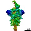
PDB-8asi:
Four subunit cytochrome b-c1 complex from Rhodobacter sphaeroides in native nanodiscs - consensus refinement in the b-b conformation
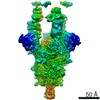
PDB-8asj:
Four subunit cytochrome b-c1 complex from Rhodobacter sphaeroides in native nanodiscs - focussed refinement in the b-c conformation

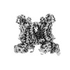
EMDB-27070:
apo form Cryo-EM structure of Campylobacter jejune ketol-acid reductoisommerase crosslinked by Glutaraldehyde
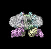
EMDB-14224:
2.8 Angstrom cryo-EM structure of the dimeric cytochrome b6f-PetP complex from Synechocystis sp. PCC 6803 with natively bound lipids and plastoquinone molecules
EMDB-15017:
3.15 Angstrom cryo-EM structure of the dimeric cytochrome b6f complex from Synechocystis sp. PCC 6803 with natively bound plastoquinone and lipid molecules.

PDB-7r0w:
2.8 Angstrom cryo-EM structure of the dimeric cytochrome b6f-PetP complex from Synechocystis sp. PCC 6803 with natively bound lipids and plastoquinone molecules

PDB-7zxy:
3.15 Angstrom cryo-EM structure of the dimeric cytochrome b6f complex from Synechocystis sp. PCC 6803 with natively bound plastoquinone and lipid molecules.

EMDB-14332:
1.58 A STRUCTURE OF HUMAN APOFERRITIN OBTAINED FROM TITAN KRIOS 2 AT eBIC, DLS UNDER COMMISSIONING SESSION CM26464-2

PDB-7r5o:
1.58 A STRUCTURE OF HUMAN APOFERRITIN OBTAINED FROM TITAN KRIOS 2 AT eBIC, DLS UNDER COMMISSIONING SESSION CM26464-2

EMDB-31081:
Subtomogram average of a meiotic triple helix from a pachytene S. cerevisiae cell.

EMDB-12679:
Cryo-EM structure (model_1a) of the RC-dLH complex from Gemmatimonas phototrophica at 2.4 A

EMDB-12680:
Cryo-EM structure (model_2a) of the RC-dLH complex from Gemmatimonas phototrophica at 2.5 A
EMDB-12681:
Cryo-EM structure of the RC-dLH complex (model_1b) from Gemmatimonas phototrophica at 2.47 A
EMDB-12682:
Cryo-EM structure (model_2b) of the RC-dLH complex from Gemmatimonas phototrophica at 2.44 A

PDB-7o0u:
Cryo-EM structure (model_1a) of the RC-dLH complex from Gemmatimonas phototrophica at 2.4 A

PDB-7o0v:
Cryo-EM structure (model_2a) of the RC-dLH complex from Gemmatimonas phototrophica at 2.5 A

PDB-7o0w:
Cryo-EM structure of the RC-dLH complex (model_1b) from Gemmatimonas phototrophica at 2.47 A

PDB-7o0x:
Cryo-EM structure (model_2b) of the RC-dLH complex from Gemmatimonas phototrophica at 2.44 A
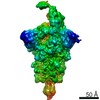
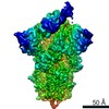
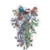
EMDB-12274:
EM structure of SARS-CoV-2 Spike glycoprotein in complex with COVOX-40 Fab
EMDB-12275:
EM structure of SARS-CoV-2 Spike glycoprotein in complex with COVOX-88 Fab
EMDB-12276:
EM structure of SARS-CoV-2 Spike glycoprotein in complex with COVOX-150 Fab
EMDB-12277:
EM structure of SARS-CoV-2 Spike glycoprotein in complex with COVOX-40 Fab
EMDB-12278:
EM structure of SARS-CoV-2 Spike glycoprotein in complex with COVOX-316 Fab
EMDB-12279:
EM structure of SARS-CoV-2 Spike glycoprotein in complex with COVOX-384 Fab
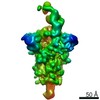
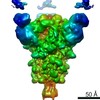
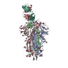
EMDB-12280:
EM structure of SARS-CoV-2 Spike glycoprotein (one RBD up) in complex with COVOX-253H55L Fab
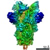
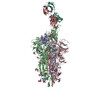
EMDB-12281:
EM structure of SARS-CoV-2 Spike glycoprotein (all RBD down) in complex with COVOX-253H55L Fab
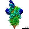
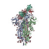
EMDB-12282:
EM structure of SARS-CoV-2 Spike glycoprotein in complex with COVOX-253H165L Fab
EMDB-12283:
EM structure of SARS-CoV-2 Spike glycoprotein (all RBD down) in complex with COVOX-159
EMDB-12284:
EM structure of SARS-CoV-2 Spike glycoprotein (one RBD up) in complex with COVOX-159
PDB-7nd3:
EM structure of SARS-CoV-2 Spike glycoprotein in complex with COVOX-40 Fab
PDB-7nd4:
EM structure of SARS-CoV-2 Spike glycoprotein in complex with COVOX-88 Fab

PDB-7nd5:
EM structure of SARS-CoV-2 Spike glycoprotein in complex with COVOX-150 Fab
PDB-7nd6:
EM structure of SARS-CoV-2 Spike glycoprotein in complex with COVOX-40 Fab
PDB-7nd7:
EM structure of SARS-CoV-2 Spike glycoprotein in complex with COVOX-316 Fab
PDB-7nd8:
EM structure of SARS-CoV-2 Spike glycoprotein in complex with COVOX-384 Fab
PDB-7nd9:
EM structure of SARS-CoV-2 Spike glycoprotein (one RBD up) in complex with COVOX-253H55L Fab
PDB-7nda:
EM structure of SARS-CoV-2 Spike glycoprotein (all RBD down) in complex with COVOX-253H55L Fab
PDB-7ndb:
EM structure of SARS-CoV-2 Spike glycoprotein in complex with COVOX-253H165L Fab
PDB-7ndc:
EM structure of SARS-CoV-2 Spike glycoprotein (all RBD down) in complex with COVOX-159

PDB-7ndd:
EM structure of SARS-CoV-2 Spike glycoprotein (one RBD up) in complex with COVOX-159
EMDB-10316:
Hepatitis B virus core protein virus-like particle displaying the antigen: extra cellular adhesion domain NadA from Neisseria meningitidis.

PDB-6tik:
Hepatitis B virus core shell--virus-like particle with NadA epitope

EMDB-10162:
Cryo-EM structure of the ClpXP1/2 protein degradation machinery
PDB-6sfw:
Cryo-EM Structure of the ClpX component of the ClpXP1/2 degradation machinery.

PDB-6sfx:
Cryo-EM structure of ClpP1/2 in the LmClpXP1/2 complex

PDB-6q81:
Structure of P-glycoprotein(ABCB1) in the post-hydrolytic state

EMDB-3882:
Thermus thermophilus PilF ATPase (apoprotein form)

EMDB-4194:
Thermus thermophilus PilF ATPase (AMPPNP-bound form)
ページ:
 wwPDBはEMDBデータモデルのバージョン3へ移行します
