 Movie
Movie Controller

[English] 日本語
 Yorodumi
- EMDB-15617: Four subunit cytochrome b-c1 complex from Rhodobacter sphaeroides... -
+ Open data

- Basic information
| Entry |  | ||||||||||||||||||
|---|---|---|---|---|---|---|---|---|---|---|---|---|---|---|---|---|---|---|---|
| Title | Four subunit cytochrome b-c1 complex from Rhodobacter sphaeroides in native nanodiscs - focussed refinement in the b-c conformation | ||||||||||||||||||
 Map data Map data | Postprocessed masked map from RELION 3.1 | ||||||||||||||||||
 Sample Sample |
| ||||||||||||||||||
 Keywords Keywords | cyt bc1 complex III membrane protein electron transport quinone cytochrome / OXIDOREDUCTASE | ||||||||||||||||||
| Function / homology |  Function and homology information Function and homology informationrespiratory chain complex III / quinol-cytochrome-c reductase / quinol-cytochrome-c reductase activity / respiratory electron transport chain / 2 iron, 2 sulfur cluster binding / electron transfer activity / oxidoreductase activity / heme binding / membrane / metal ion binding / plasma membrane Similarity search - Function | ||||||||||||||||||
| Biological species |  Cereibacter sphaeroides (bacteria) / Cereibacter sphaeroides 2.4.1 (bacteria) Cereibacter sphaeroides (bacteria) / Cereibacter sphaeroides 2.4.1 (bacteria) | ||||||||||||||||||
| Method | single particle reconstruction / cryo EM / Resolution: 3.75 Å | ||||||||||||||||||
 Authors Authors | Swainsbury DJK / Hawkings FR / Martin EC / Musial S / Salisbury JH / Jackson PJ / Farmer DA / Johnson MP / Siebert CA / Hitchcock A / Hunter CN | ||||||||||||||||||
| Funding support | European Union,  United Kingdom, 5 items United Kingdom, 5 items
| ||||||||||||||||||
 Citation Citation | Journal: Proc Natl Acad Sci U S A / Year: 2023 Title: Cryo-EM structure of the four-subunit cytochrome complex in styrene maleic acid nanodiscs. Authors: David J K Swainsbury / Frederick R Hawkings / Elizabeth C Martin / Sabina Musiał / Jack H Salisbury / Philip J Jackson / David A Farmer / Matthew P Johnson / C Alistair Siebert / Andrew ...Authors: David J K Swainsbury / Frederick R Hawkings / Elizabeth C Martin / Sabina Musiał / Jack H Salisbury / Philip J Jackson / David A Farmer / Matthew P Johnson / C Alistair Siebert / Andrew Hitchcock / C Neil Hunter / Abstract: Cytochrome complexes are ubiquinol:cytochrome oxidoreductases, and as such, they are centrally important components of respiratory and photosynthetic electron transfer chains in many species of ...Cytochrome complexes are ubiquinol:cytochrome oxidoreductases, and as such, they are centrally important components of respiratory and photosynthetic electron transfer chains in many species of bacteria and in mitochondria. The minimal complex has three catalytic components, which are cytochrome , cytochrome , and the Rieske iron-sulfur subunit, but the function of mitochondrial cytochrome complexes is modified by up to eight supernumerary subunits. The cytochrome complex from the purple phototrophic bacterium has a single supernumerary subunit called subunit IV, which is absent from current structures of the complex. In this work we use the styrene-maleic acid copolymer to purify the cytochrome complex in native lipid nanodiscs, which retains the labile subunit IV, annular lipids, and natively bound quinones. The catalytic activity of the four-subunit cytochrome complex is threefold higher than that of the complex lacking subunit IV. To understand the role of subunit IV, we determined the structure of the four-subunit complex at 2.9 Å using single particle cryogenic electron microscopy. The structure shows the position of the transmembrane domain of subunit IV, which lies across the transmembrane helices of the Rieske and cytochrome subunits. We observe a quinone at the Q quinone-binding site and show that occupancy of this site is linked to conformational changes in the Rieske head domain during catalysis. Twelve lipids were structurally resolved, making contacts with the Rieske and cytochrome subunits, with some spanning both of the two monomers that make up the dimeric complex. | ||||||||||||||||||
| History |
|
- Structure visualization
| Supplemental images |
|---|

- Downloads & links
-EMDB archive
| Map data | emd_15617.map.gz | 4.5 MB | EMDB map data format | |
|---|---|---|---|---|
| Header (meta data) | emd-15617-v30.xmlemd-15617.xml | 32.3 KB 32.3 KB | Display Display | EMDB header |
| FSC (resolution estimation) | emd_15617_fsc.xml | 7.8 KB | Display | FSC data file |
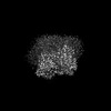
| Images | 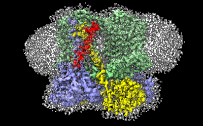 emd_15617.png emd_15617.png | 140.4 KB | ||
| Filedesc metadata | emd-15617.cif.gz | 8.6 KB | ||
| Others | emd_15617_additional_1.map.gzemd_15617_half_map_1.map.gzemd_15617_half_map_2.map.gz | 5.2 MB 31.4 MB 31.4 MB | ||
| Archive directory |  http://ftp.pdbj.org/pub/emdb/structures/EMD-15617ftp://ftp.pdbj.org/pub/emdb/structures/EMD-15617 http://ftp.pdbj.org/pub/emdb/structures/EMD-15617ftp://ftp.pdbj.org/pub/emdb/structures/EMD-15617 | HTTPS FTP |
-Related structure data
| Related structure data | 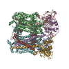 8asjMC 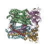 8asiC M: atomic model generated by this map C: citing same article ( |
|---|---|
| Similar structure data |
-Links
| EMDB pages | EMDB (EBI/PDBe) / EMDataResource |
|---|---|
| Related items in Molecule of the Month |


-Map
| File | Download / File: emd_15617.map.gz / Format: CCP4 / Size: 40.6 MB / Type: IMAGE STORED AS FLOATING POINT NUMBER (4 BYTES) | ||||||||||||||||||||||||||||||||||||
|---|---|---|---|---|---|---|---|---|---|---|---|---|---|---|---|---|---|---|---|---|---|---|---|---|---|---|---|---|---|---|---|---|---|---|---|---|---|
| Annotation | Postprocessed masked map from RELION 3.1 | ||||||||||||||||||||||||||||||||||||
| Projections & slices | Image control
Images are generated by Spider. | ||||||||||||||||||||||||||||||||||||
| Voxel size | X=Y=Z: 1.1718 Å | ||||||||||||||||||||||||||||||||||||
| Density |
| ||||||||||||||||||||||||||||||||||||
| Symmetry | Space group: 1 | ||||||||||||||||||||||||||||||||||||
| Details | EMDB XML:
|
 Z (Sec.)
 Y (Row.)
 X (Col.)


-Supplemental data
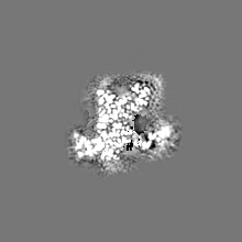
-Additional map: Locally sharpened map using LocScale
| File | emd_15617_additional_1.map | ||||||||||||
|---|---|---|---|---|---|---|---|---|---|---|---|---|---|
| Annotation | Locally sharpened map using LocScale | ||||||||||||
| Projections & Slices |
| ||||||||||||
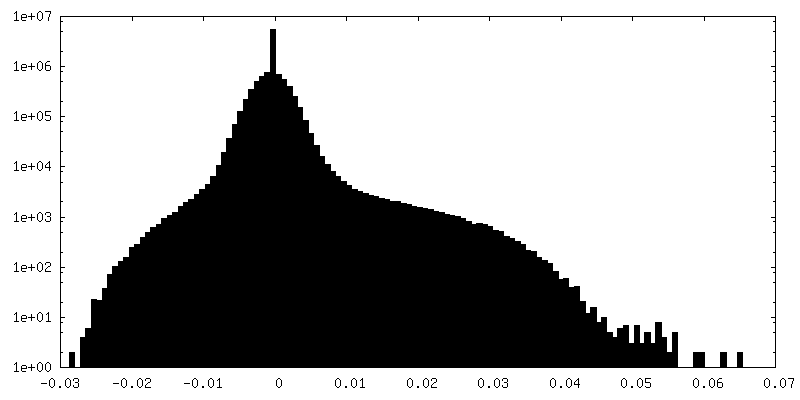
| Density Histograms |


-Half map: Half map 1
| File | emd_15617_half_map_1.map | ||||||||||||
|---|---|---|---|---|---|---|---|---|---|---|---|---|---|
| Annotation | Half map 1 | ||||||||||||
| Projections & Slices |
| ||||||||||||
| Density Histograms |


-Half map: Half map 2
| File | emd_15617_half_map_2.map | ||||||||||||
|---|---|---|---|---|---|---|---|---|---|---|---|---|---|
| Annotation | Half map 2 | ||||||||||||
| Projections & Slices |
| ||||||||||||
| Density Histograms |


- Sample components
+Entire : Four subunit cytochrome b-c1 complex from Rhodobacter sphaeroides
+Supramolecule #1: Four subunit cytochrome b-c1 complex from Rhodobacter sphaeroides
+Macromolecule #1: Ubiquinol-cytochrome c reductase iron-sulfur subunit
+Macromolecule #2: Cytochrome b
+Macromolecule #3: Cytochrome c1
+Macromolecule #4: Cytochrome b-c1 subunit IV
+Macromolecule #5: FE2/S2 (INORGANIC) CLUSTER

+Macromolecule #6: 1,2-dioleoyl-sn-glycero-3-phosphoethanolamine

+Macromolecule #7: PROTOPORPHYRIN IX CONTAINING FE

+Macromolecule #8: HEME C

+Macromolecule #9: UBIQUINONE-10

-Experimental details
-Structure determination
| Method | cryo EM |
|---|---|
 Processing Processing | single particle reconstruction |
| Aggregation state | particle |
-Sample preparation
| Buffer | pH: 8 Component:
Details: Solutions were freshly prepared from concentrated stock solutions. | |||||||||
|---|---|---|---|---|---|---|---|---|---|---|
| Grid | Model: Quantifoil R1.2/1.3 / Material: COPPER / Mesh: 300 / Support film - Material: CARBON / Support film - topology: HOLEY ARRAY | |||||||||
| Vitrification | Cryogen name: ETHANE / Chamber humidity: 80 % / Chamber temperature: 298 K / Instrument: LEICA EM GP Details: 15 ul of sample was applied to the grid, incubated for 30 s, blotted for 4 s then plunged in liquid ethane.. | |||||||||
| Details | Monodisperse particles consisting of four-subunit cyt b-c1 solubilised in styrene maleic acid nanodiscs |
- Electron microscopy
| Microscope | FEI TITAN KRIOS |
|---|---|
| Specialist optics | Energy filter - Slit width: 20 eV |
| Image recording | Film or detector model: GATAN K3 (6k x 4k) / Digitization - Dimensions - Width: 5760 pixel / Digitization - Dimensions - Height: 4092 pixel / Number grids imaged: 1 / Number real images: 15867 / Average exposure time: 1.13 sec. / Average electron dose: 1.0 e/Å2 |
| Electron beam | Acceleration voltage: 300 kV / Electron source:  FIELD EMISSION GUN FIELD EMISSION GUN |
| Electron optics | C2 aperture diameter: 50.0 µm / Illumination mode: FLOOD BEAM / Imaging mode: BRIGHT FIELD / Cs: 2.7 mm / Nominal defocus max: 2.0 µm / Nominal defocus min: 0.8 µm / Nominal magnification: 130000 |
| Sample stage | Specimen holder model: FEI TITAN KRIOS AUTOGRID HOLDER / Cooling holder cryogen: NITROGEN |
| Experimental equipment |  Model: Titan Krios / Image courtesy: FEI Company |
+Image processing

-Atomic model buiding 1
| Refinement | Space: REAL |
|---|---|
| Output model | PDB-8asj: |
