 Movie
Movie Controller

[English] 日本語
 Yorodumi
- PDB-8asi: Four subunit cytochrome b-c1 complex from Rhodobacter sphaeroides... -
+ Open data

- Basic information
| Entry | Database: PDB / ID: 8asi | |||||||||||||||||||||||||||||||||||||||||||||
|---|---|---|---|---|---|---|---|---|---|---|---|---|---|---|---|---|---|---|---|---|---|---|---|---|---|---|---|---|---|---|---|---|---|---|---|---|---|---|---|---|---|---|---|---|---|---|
| Title | Four subunit cytochrome b-c1 complex from Rhodobacter sphaeroides in native nanodiscs - consensus refinement in the b-b conformation | |||||||||||||||||||||||||||||||||||||||||||||
 Components Components |
| |||||||||||||||||||||||||||||||||||||||||||||
 Keywords Keywords | OXIDOREDUCTASE / cyt bc1 complex III membrane protein electron transport quinone cytochrome | |||||||||||||||||||||||||||||||||||||||||||||
| Function / homology |  Function and homology information Function and homology informationrespiratory chain complex III / quinol-cytochrome-c reductase / quinol-cytochrome-c reductase activity / respiratory electron transport chain / 2 iron, 2 sulfur cluster binding / electron transfer activity / oxidoreductase activity / heme binding / membrane / metal ion binding / plasma membrane Similarity search - Function | |||||||||||||||||||||||||||||||||||||||||||||
| Biological species |  Cereibacter sphaeroides 2.4.1 (bacteria) Cereibacter sphaeroides 2.4.1 (bacteria) | |||||||||||||||||||||||||||||||||||||||||||||
| Method | ELECTRON MICROSCOPY / single particle reconstruction / cryo EM / Resolution: 2.9 Å | |||||||||||||||||||||||||||||||||||||||||||||
 Authors Authors | Swainsbury, D.J.K. / Hawkings, F.R. / Martin, E.C. / Musial, S. / Salisbury, J.H. / Jackson, P.J. / Farmer, D.A. / Johnson, M.P. / Siebert, C.A. / Hitchcock, A. / Hunter, C.N. | |||||||||||||||||||||||||||||||||||||||||||||
| Funding support | European Union,  United Kingdom, 5items United Kingdom, 5items
| |||||||||||||||||||||||||||||||||||||||||||||
 Citation Citation | Journal: Proc Natl Acad Sci U S A / Year: 2023 Title: Cryo-EM structure of the four-subunit cytochrome complex in styrene maleic acid nanodiscs. Authors: David J K Swainsbury / Frederick R Hawkings / Elizabeth C Martin / Sabina Musiał / Jack H Salisbury / Philip J Jackson / David A Farmer / Matthew P Johnson / C Alistair Siebert / Andrew ...Authors: David J K Swainsbury / Frederick R Hawkings / Elizabeth C Martin / Sabina Musiał / Jack H Salisbury / Philip J Jackson / David A Farmer / Matthew P Johnson / C Alistair Siebert / Andrew Hitchcock / C Neil Hunter / Abstract: Cytochrome complexes are ubiquinol:cytochrome oxidoreductases, and as such, they are centrally important components of respiratory and photosynthetic electron transfer chains in many species of ...Cytochrome complexes are ubiquinol:cytochrome oxidoreductases, and as such, they are centrally important components of respiratory and photosynthetic electron transfer chains in many species of bacteria and in mitochondria. The minimal complex has three catalytic components, which are cytochrome , cytochrome , and the Rieske iron-sulfur subunit, but the function of mitochondrial cytochrome complexes is modified by up to eight supernumerary subunits. The cytochrome complex from the purple phototrophic bacterium has a single supernumerary subunit called subunit IV, which is absent from current structures of the complex. In this work we use the styrene-maleic acid copolymer to purify the cytochrome complex in native lipid nanodiscs, which retains the labile subunit IV, annular lipids, and natively bound quinones. The catalytic activity of the four-subunit cytochrome complex is threefold higher than that of the complex lacking subunit IV. To understand the role of subunit IV, we determined the structure of the four-subunit complex at 2.9 Å using single particle cryogenic electron microscopy. The structure shows the position of the transmembrane domain of subunit IV, which lies across the transmembrane helices of the Rieske and cytochrome subunits. We observe a quinone at the Q quinone-binding site and show that occupancy of this site is linked to conformational changes in the Rieske head domain during catalysis. Twelve lipids were structurally resolved, making contacts with the Rieske and cytochrome subunits, with some spanning both of the two monomers that make up the dimeric complex. | |||||||||||||||||||||||||||||||||||||||||||||
| History |
|
- Structure visualization
| Structure viewer | Molecule: MolmilJmol/JSmol |
|---|
- Downloads & links
-Download
| PDBx/mmCIF format | 8asi.cif.gz | 342.6 KB | Display | PDBx/mmCIF format |
|---|---|---|---|---|
| PDB format | pdb8asi.ent.gz | 272.3 KB | Display | PDB format |
| PDBx/mmJSON format | 8asi.json.gz | Tree view | PDBx/mmJSON format | |
| Others |  Other downloads Other downloads |
-Validation report
| Arichive directory | https://data.pdbj.org/pub/pdb/validation_reports/as/8asiftp://data.pdbj.org/pub/pdb/validation_reports/as/8asi | HTTPS FTP |
|---|
-Related structure data
| Related structure data | 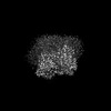 15616MC 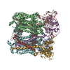 8asjC M: map data used to model this data C: citing same article ( |
|---|---|
| Similar structure data |
-Links
 PDBj
PDBj


- Assembly
| Deposited unit | 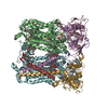
|
|---|---|
| 1 |
|
-Components
-Protein , 4 types, 8 molecules AEBFCGDH
| #1: Protein | Mass: 19928.375 Da / Num. of mol.: 2 / Source method: isolated from a natural source / Source: (natural) Cereibacter sphaeroides 2.4.1 (bacteria)Strain: ATCC 17023 / DSM 158 / JCM 6121 / CCUG 31486 / LMG 2827 / NBRC 12203 / NCIMB 8253 / ATH 2.4.1. References: UniProt: Q3IY09, quinol-cytochrome-c reductase #2: Protein | Mass: 50087.422 Da / Num. of mol.: 2 / Source method: isolated from a natural source / Source: (natural) Cereibacter sphaeroides 2.4.1 (bacteria)Strain: ATCC 17023 / DSM 158 / JCM 6121 / CCUG 31486 / LMG 2827 / NBRC 12203 / NCIMB 8253 / ATH 2.4.1. References: UniProt: Q3IY10 #3: Protein | Mass: 30661.664 Da / Num. of mol.: 2 / Source method: isolated from a natural source / Source: (natural) Cereibacter sphaeroides 2.4.1 (bacteria)Strain: ATCC 17023 / DSM 158 / JCM 6121 / CCUG 31486 / LMG 2827 / NBRC 12203 / NCIMB 8253 / ATH 2.4.1. References: UniProt: Q3IY11 #4: Protein | Mass: 14415.301 Da / Num. of mol.: 2 / Source method: isolated from a natural source / Source: (natural) Cereibacter sphaeroides 2.4.1 (bacteria)Strain: ATCC 17023 / DSM 158 / JCM 6121 / CCUG 31486 / LMG 2827 / NBRC 12203 / NCIMB 8253 / ATH 2.4.1. References: UniProt: Q3J2Z2 |
|---|
-Non-polymers , 5 types, 21 molecules 


| #5: Chemical |  Mass: 175.820 Da / Num. of mol.: 2 / Source method: obtained synthetically / Formula: Fe2S2 / Feature type: SUBJECT OF INVESTIGATION Mass: 175.820 Da / Num. of mol.: 2 / Source method: obtained synthetically / Formula: Fe2S2 / Feature type: SUBJECT OF INVESTIGATION#6: Chemical | ChemComp-PEE /  Mass: 744.034 Da / Num. of mol.: 12 / Source method: obtained synthetically / Formula: C41H78NO8P / Feature type: SUBJECT OF INVESTIGATION / Comment: DOPE, phospholipid*YM Mass: 744.034 Da / Num. of mol.: 12 / Source method: obtained synthetically / Formula: C41H78NO8P / Feature type: SUBJECT OF INVESTIGATION / Comment: DOPE, phospholipid*YM#7: Chemical | ChemComp-HEM /  Mass: 616.487 Da / Num. of mol.: 4 / Source method: obtained synthetically / Formula: C34H32FeN4O4 / Feature type: SUBJECT OF INVESTIGATION Mass: 616.487 Da / Num. of mol.: 4 / Source method: obtained synthetically / Formula: C34H32FeN4O4 / Feature type: SUBJECT OF INVESTIGATION#8: Chemical |  Mass: 618.503 Da / Num. of mol.: 2 / Source method: obtained synthetically / Formula: C34H34FeN4O4 / Feature type: SUBJECT OF INVESTIGATION Mass: 618.503 Da / Num. of mol.: 2 / Source method: obtained synthetically / Formula: C34H34FeN4O4 / Feature type: SUBJECT OF INVESTIGATION#9: Chemical | ChemComp-U10 / |  Mass: 863.343 Da / Num. of mol.: 1 / Source method: obtained synthetically / Formula: C59H90O4 / Feature type: SUBJECT OF INVESTIGATION Mass: 863.343 Da / Num. of mol.: 1 / Source method: obtained synthetically / Formula: C59H90O4 / Feature type: SUBJECT OF INVESTIGATION |
|---|
-Details
| Has ligand of interest | Y |
|---|---|
| Has protein modification | Y |
-Experimental details
-Experiment
| Experiment | Method: ELECTRON MICROSCOPY |
|---|---|
| EM experiment | Aggregation state: PARTICLE / 3D reconstruction method: single particle reconstruction |
- Sample preparation
| Component | Name: Four subunit cytochrome b-c1 complex from Rhodobacter sphaeroides Type: COMPLEX Details: Four subunit cytochrome b-c1 complex from Rhodobacter sphaeroides purified in SMA copolymer lipid nanodiscs Entity ID: #1-#4 / Source: NATURAL | |||||||||||||||
|---|---|---|---|---|---|---|---|---|---|---|---|---|---|---|---|---|
| Molecular weight | Experimental value: NO | |||||||||||||||
| Source (natural) | Organism: Cereibacter sphaeroides (bacteria) / Strain: 2.4.1 | |||||||||||||||
| Buffer solution | pH: 8 Details: Solutions were freshly prepared from concentrated stock solutions. | |||||||||||||||
| Buffer component |
| |||||||||||||||
| Specimen | Embedding applied: NO / Shadowing applied: NO / Staining applied: NO / Vitrification applied: YES Details: Monodisperse particles consisting of four-subunit cyt b-c1 solubilised in styrene maleic acid nanodiscs | |||||||||||||||
| Specimen support | Grid material: COPPER / Grid mesh size: 300 divisions/in. / Grid type: Quantifoil R1.2/1.3 | |||||||||||||||
| Vitrification | Instrument: LEICA EM GP / Cryogen name: ETHANE / Humidity: 80 % / Chamber temperature: 298 K Details: 15 ul of sample was applied to the grid, incubated for 30 s, blotted for 4 s then plunged in liquid ethane. |
- Electron microscopy imaging
| Experimental equipment |  Model: Titan Krios / Image courtesy: FEI Company |
|---|---|
| Microscopy | Model: FEI TITAN KRIOS |
| Electron gun | Electron source:  FIELD EMISSION GUN / Accelerating voltage: 300 kV / Illumination mode: FLOOD BEAM FIELD EMISSION GUN / Accelerating voltage: 300 kV / Illumination mode: FLOOD BEAM |
| Electron lens | Mode: BRIGHT FIELD / Nominal magnification: 130000 X / Nominal defocus max: 2000 nm / Nominal defocus min: 800 nm / Cs: 2.7 mm / C2 aperture diameter: 50 µm / Alignment procedure: COMA FREE |
| Specimen holder | Cryogen: NITROGEN / Specimen holder model: FEI TITAN KRIOS AUTOGRID HOLDER |
| Image recording | Average exposure time: 1.13 sec. / Electron dose: 1 e/Å2 / Film or detector model: GATAN K3 (6k x 4k) / Num. of grids imaged: 1 / Num. of real images: 15867 |
| EM imaging optics | Energyfilter slit width: 20 eV |
| Image scans | Width: 5760 / Height: 4092 |
- Processing
| Software | Name: PHENIX / Version: 1.19.2_4158: / Classification: refinement | ||||||||||||||||||||||||||||||||||||||||
|---|---|---|---|---|---|---|---|---|---|---|---|---|---|---|---|---|---|---|---|---|---|---|---|---|---|---|---|---|---|---|---|---|---|---|---|---|---|---|---|---|---|
| EM software |
| ||||||||||||||||||||||||||||||||||||||||
| Image processing | Details: Images were motion corrected using Motiocorr 2 within RELION using 5 x 5 patches. | ||||||||||||||||||||||||||||||||||||||||
| CTF correction | Type: PHASE FLIPPING AND AMPLITUDE CORRECTION | ||||||||||||||||||||||||||||||||||||||||
| Particle selection | Num. of particles selected: 4060135 Details: Particles were picked using crYOLO 1.7.5 using a model trained on this dataset | ||||||||||||||||||||||||||||||||||||||||
| Symmetry | Point symmetry: C1 (asymmetric) | ||||||||||||||||||||||||||||||||||||||||
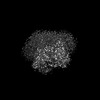
| 3D reconstruction | Resolution: 2.9 Å / Resolution method: FSC 0.143 CUT-OFF / Num. of particles: 282636 / Algorithm: FOURIER SPACE / Num. of class averages: 1 / Symmetry type: POINT | ||||||||||||||||||||||||||||||||||||||||
| Atomic model building | Space: REAL | ||||||||||||||||||||||||||||||||||||||||
| Refinement | Cross valid method: NONE Stereochemistry target values: GeoStd + Monomer Library + CDL v1.2 | ||||||||||||||||||||||||||||||||||||||||
| Displacement parameters | Biso mean: 74.72 Å2 | ||||||||||||||||||||||||||||||||||||||||
| Refine LS restraints |
|
