Biotechnology and Biological Sciences Research Council
BRIC PhD, BBSRC Reference BB/K02034X/1
United Kingdom
Citation
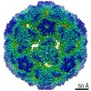
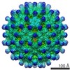
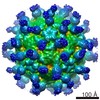
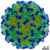
Journal: Vaccine / Year: 2020 Title: An assessment of the use of Hepatitis B Virus core protein virus-like particles to display heterologous antigens from Neisseria meningitidis. Authors: Sebastian Aston-Deaville / Emil Carlsson / Muhammad Saleem / Angela Thistlethwaite / Hannah Chan / Sunil Maharjan / Alessandra Facchetti / Ian M Feavers / C Alistair Siebert / Richard F ...Authors: Sebastian Aston-Deaville / Emil Carlsson / Muhammad Saleem / Angela Thistlethwaite / Hannah Chan / Sunil Maharjan / Alessandra Facchetti / Ian M Feavers / C Alistair Siebert / Richard F Collins / Alan Roseman / Jeremy P Derrick / Abstract: Neisseria meningitidis is the causative agent of meningococcal meningitis and sepsis and remains a significant public health problem in many countries. Efforts to develop a comprehensive vaccine ...Neisseria meningitidis is the causative agent of meningococcal meningitis and sepsis and remains a significant public health problem in many countries. Efforts to develop a comprehensive vaccine against serogroup B meningococci have focused on the use of surface-exposed outer membrane proteins. Here we report the use of virus-like particles derived from the core protein of Hepatitis B Virus, HBc, to incorporate antigen domains derived from Factor H binding protein (FHbp) and the adhesin NadA. The extracellular domain of NadA was inserted into the major immunodominant region of HBc, and the C-terminal domain of FHbp at the C-terminus (CFHbp), creating a single polypeptide chain 3.7-fold larger than native HBc. Remarkably, cryoelectron microscopy revealed that the construct formed assemblies that were able to incorporate both antigens with minimal structural changes to native HBc. Electron density was weak for NadA and absent for CFHbp, partly attributable to domain flexibility. Following immunization of mice, three HBc fusions (CFHbp or NadA alone, NadA + CFHbp) were able to induce production of IgG1, IgG2a and IgG2b antibodies reactive against their respective antigens at dilutions in excess of 1:18,000. However, only HBc fusions containing NadA elicited the production of antibodies with serum bactericidal activity. It is hypothesized that this improved immune response is attributable to the adoption of a more native-like folding of crucial conformational epitopes of NadA within the chimeric VLP. This work demonstrates that HBc can incorporate insertions of large antigen domains but that maintenance of their three-dimensional structure is likely to be critical in obtaining a protective response.
History
Deposition
Sep 18, 2019
-
Header (metadata) release
Jul 29, 2020
-
Map release
Jul 29, 2020
-
Update
Nov 25, 2020
-
Current status
Nov 25, 2020
Processing site: PDBe / Status: Released
-
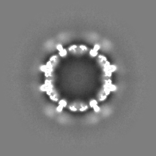
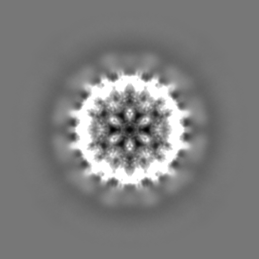
Structure visualization
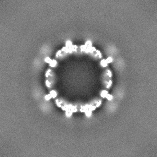
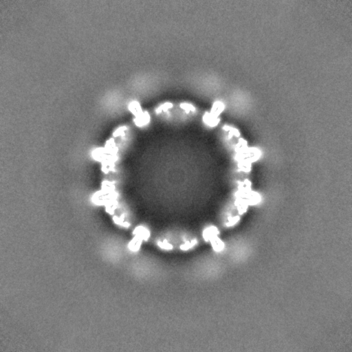
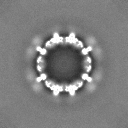
Movie
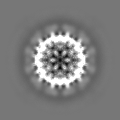
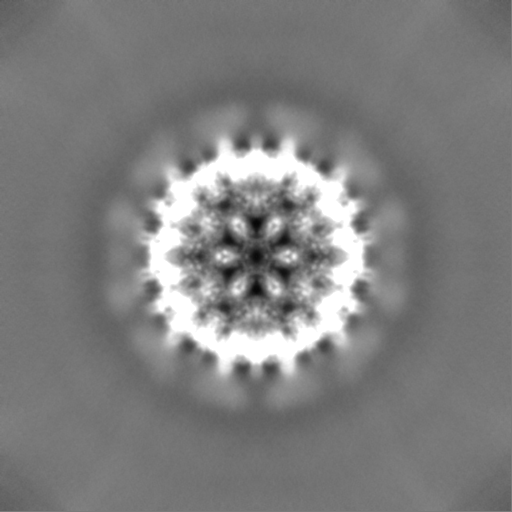
Surface view with section colored by density value
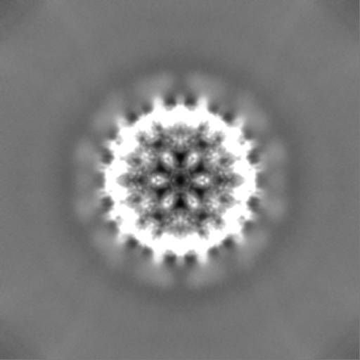
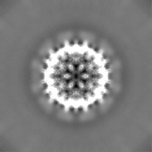
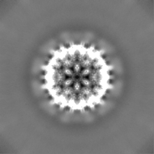
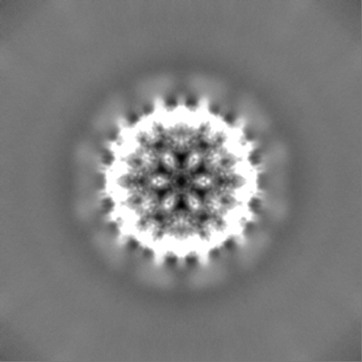
Supramolecule #1: VLP fusion sequence expressed in E.coli.
Supramolecule
Name: VLP fusion sequence expressed in E.coli. / type: complex / ID: 1 / Parent: 0 / Macromolecule list: all Details: HBcS-NadA-CFHbp Fusion protein with linker regions. HBc - Ordered core of VLP: from HBV core protein (NCBI taxonomy ID 10407). NadA - Partially ordered surface displayed domain: ...Details: HBcS-NadA-CFHbp Fusion protein with linker regions. HBc - Ordered core of VLP: from HBV core protein (NCBI taxonomy ID 10407). NadA - Partially ordered surface displayed domain: extracellular domain of NadA from Neisseria meningitidis (NCBI taxonomy ID 487). CFHbp - Unresolved/disordered component : C-terminal domain of factor H binding protein from Neisseria meningitidis (NCBI taxonomy ID 487).
Cryogen name: ETHANE / Chamber humidity: 90 % / Chamber temperature: 295 K / Instrument: FEI VITROBOT MARK IV Details: 3 ul applied for 30s at room temperature, then 4-5 s blot..
-
Electron microscopy
Microscope
FEI TITAN KRIOS
Image recording
Film or detector model: FEI FALCON III (4k x 4k) / Detector mode: INTEGRATING / Digitization - Dimensions - Width: 4096 pixel / Digitization - Dimensions - Height: 4096 pixel / Digitization - Sampling interval: 14.0 µm / Digitization - Frames/image: 3-36 / Number grids imaged: 1 / Number real images: 2367 / Average exposure time: 4.0 sec. / Average electron dose: 79.0 e/Å2
Electron beam
Acceleration voltage: 300 kV / Electron source: FIELD EMISSION GUN
Number selected: 10119 Details: Initially ~44,000 particles were initially picked and extracted in EMAN. Approx 12,000 were shell fragments or incomplete. Others were intact but lower resolution, so damaged or affected by ...Details: Initially ~44,000 particles were initially picked and extracted in EMAN. Approx 12,000 were shell fragments or incomplete. Others were intact but lower resolution, so damaged or affected by charging, or other issue. After filtering/cleaning by EMAN2 2D classification 10119 particles were taken forwards for initial 3D reconstruction in EMAN2. This set of 10119 was then 2D cleaned by CryoSPARC to give 9145 particles.
CTF correction
Software - Name: EMAN2 (ver. 2.01) / Software - details: then CTF applied by CryoSPARC
Final reconstruction
Applied symmetry - Point group: I (icosahedral) / Resolution.type: BY AUTHOR / Resolution: 3.4 Å / Resolution method: FSC 0.143 CUT-OFF / Software - Name: cryoSPARC (ver. v0.65) / Number images used: 8598
Initial angle assignment
Type: OTHER / Software - Name: cryoSPARC (ver. v0.65)
Final angle assignment
Type: MAXIMUM LIKELIHOOD / Software - Name: cryoSPARC (ver. v0.65)
Final 3D classification
Number classes: 3 / Software - Name: cryoSPARC (ver. v0.65) / Details: model #0 94.2 % mode l#1 4.6 % model #2 1.2%
+
About Yorodumi
-
News
-
Feb 9, 2022. New format data for meta-information of EMDB entries
New format data for meta-information of EMDB entries
Version 3 of the EMDB header file is now the official format.
The previous official version 1.9 will be removed from the archive.
In the structure databanks used in Yorodumi, some data are registered as the other names, "COVID-19 virus" and "2019-nCoV". Here are the details of the virus and the list of structure data.
Jan 31, 2019. EMDB accession codes are about to change! (news from PDBe EMDB page)
EMDB accession codes are about to change! (news from PDBe EMDB page)
The allocation of 4 digits for EMDB accession codes will soon come to an end. Whilst these codes will remain in use, new EMDB accession codes will include an additional digit and will expand incrementally as the available range of codes is exhausted. The current 4-digit format prefixed with “EMD-” (i.e. EMD-XXXX) will advance to a 5-digit format (i.e. EMD-XXXXX), and so on. It is currently estimated that the 4-digit codes will be depleted around Spring 2019, at which point the 5-digit format will come into force.
The EM Navigator/Yorodumi systems omit the EMD- prefix.
Related info.:Q: What is EMD? / ID/Accession-code notation in Yorodumi/EM Navigator
Yorodumi is a browser for structure data from EMDB, PDB, SASBDB, etc.
This page is also the successor to EM Navigator detail page, and also detail information page/front-end page for Omokage search.
The word "yorodu" (or yorozu) is an old Japanese word meaning "ten thousand". "mi" (miru) is to see.
Related info.:EMDB / PDB / SASBDB / Comparison of 3 databanks / Yorodumi Search / Aug 31, 2016. New EM Navigator & Yorodumi / Yorodumi Papers / Jmol/JSmol / Function and homology information / Changes in new EM Navigator and Yorodumi
 Movie
Movie Controller

 Yorodumi
 Open data
 Basic information
 Map data
 Sample
 Function and homology information
 Neisseria meningitidis (bacteria)
 Authors
 United Kingdom, 1 items
  Citation
 Structure visualization
 Downloads & links
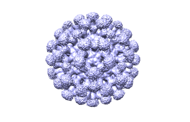 emd_10316.png
 http://ftp.pdbj.org/pub/emdb/structures/EMD-10316


 Z (Sec.)
 Y (Row.)
 X (Col.)


 Sample components
 Processing
 Electron microscopy
 FIELD EMISSION GUN
