 Movie
Movie Controller

[English] 日本語
 Yorodumi
- PDB-6tik: Hepatitis B virus core shell--virus-like particle with NadA epitope -
+ Open data

- Basic information
| Entry | Database: PDB / ID: 6tik | ||||||
|---|---|---|---|---|---|---|---|
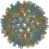
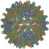
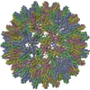
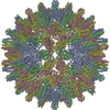
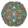
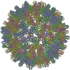
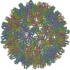
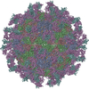
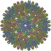
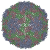
| Title | Hepatitis B virus core shell--virus-like particle with NadA epitope | ||||||
 Components Components | Capsid protein,Putative adhesin/invasin,Capsid protein,Factor H-binding protein | ||||||
 Keywords Keywords | VIRUS LIKE PARTICLE / Virus-like particle / VLP / antigen / NadA / Neisseria meningitidis / HBV / HBC / factor H binding protein | ||||||
| Function / homology |  Function and homology information Function and homology informationmicrotubule-dependent intracellular transport of viral material towards nucleus / T=4 icosahedral viral capsid / cell outer membrane / viral penetration into host nucleus / host cell / host cell cytoplasm / viral envelope / symbiont entry into host cell / structural molecule activity / cell surface ...microtubule-dependent intracellular transport of viral material towards nucleus / T=4 icosahedral viral capsid / cell outer membrane / viral penetration into host nucleus / host cell / host cell cytoplasm / viral envelope / symbiont entry into host cell / structural molecule activity / cell surface / DNA binding / RNA binding / membrane Similarity search - Function | ||||||
| Biological species |   Hepatitis B virus Hepatitis B virus Neisseria meningitidis (bacteria) Neisseria meningitidis (bacteria) | ||||||
| Method | ELECTRON MICROSCOPY / single particle reconstruction / cryo EM / Resolution: 3.4 Å | ||||||
 Authors Authors | Roseman, A.M. / Colllins, R.F. / Derrick, J.P. | ||||||
| Funding support |  United Kingdom, 1items United Kingdom, 1items
| ||||||
 Citation Citation | Journal: Vaccine / Year: 2020 Title: An assessment of the use of Hepatitis B Virus core protein virus-like particles to display heterologous antigens from Neisseria meningitidis. Authors: Sebastian Aston-Deaville / Emil Carlsson / Muhammad Saleem / Angela Thistlethwaite / Hannah Chan / Sunil Maharjan / Alessandra Facchetti / Ian M Feavers / C Alistair Siebert / Richard F ...Authors: Sebastian Aston-Deaville / Emil Carlsson / Muhammad Saleem / Angela Thistlethwaite / Hannah Chan / Sunil Maharjan / Alessandra Facchetti / Ian M Feavers / C Alistair Siebert / Richard F Collins / Alan Roseman / Jeremy P Derrick / Abstract: Neisseria meningitidis is the causative agent of meningococcal meningitis and sepsis and remains a significant public health problem in many countries. Efforts to develop a comprehensive vaccine ...Neisseria meningitidis is the causative agent of meningococcal meningitis and sepsis and remains a significant public health problem in many countries. Efforts to develop a comprehensive vaccine against serogroup B meningococci have focused on the use of surface-exposed outer membrane proteins. Here we report the use of virus-like particles derived from the core protein of Hepatitis B Virus, HBc, to incorporate antigen domains derived from Factor H binding protein (FHbp) and the adhesin NadA. The extracellular domain of NadA was inserted into the major immunodominant region of HBc, and the C-terminal domain of FHbp at the C-terminus (CFHbp), creating a single polypeptide chain 3.7-fold larger than native HBc. Remarkably, cryoelectron microscopy revealed that the construct formed assemblies that were able to incorporate both antigens with minimal structural changes to native HBc. Electron density was weak for NadA and absent for CFHbp, partly attributable to domain flexibility. Following immunization of mice, three HBc fusions (CFHbp or NadA alone, NadA + CFHbp) were able to induce production of IgG1, IgG2a and IgG2b antibodies reactive against their respective antigens at dilutions in excess of 1:18,000. However, only HBc fusions containing NadA elicited the production of antibodies with serum bactericidal activity. It is hypothesized that this improved immune response is attributable to the adoption of a more native-like folding of crucial conformational epitopes of NadA within the chimeric VLP. This work demonstrates that HBc can incorporate insertions of large antigen domains but that maintenance of their three-dimensional structure is likely to be critical in obtaining a protective response. | ||||||
| History |
|
- Structure visualization
| Movie |
Movie viewer |
|---|---|
| Structure viewer | Molecule: MolmilJmol/JSmol |
- Downloads & links
-Download
| PDBx/mmCIF format | 6tik.cif.gz | 138.2 KB | Display | PDBx/mmCIF format |
|---|---|---|---|---|
| PDB format | pdb6tik.ent.gz | 92.2 KB | Display | PDB format |
| PDBx/mmJSON format | 6tik.json.gz | Tree view | PDBx/mmJSON format | |
| Others |  Other downloads Other downloads |
-Validation report
| Arichive directory | https://data.pdbj.org/pub/pdb/validation_reports/ti/6tikftp://data.pdbj.org/pub/pdb/validation_reports/ti/6tik | HTTPS FTP |
|---|
-Related structure data
| Related structure data | 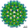 10316MC M: map data used to model this data C: citing same article ( |
|---|---|
| Similar structure data |
-Links
 PDBj
PDBj


- Assembly
| Deposited unit | 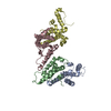
|
|---|---|
| 1 | x 60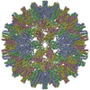
|
-Components
| #1: Protein | Mass: 62116.750 Da / Num. of mol.: 4 Source method: isolated from a genetically manipulated source Details: HBcS-NadA-CFHbp VLP fusion sequence expressed in E.coli. HBc - core shell from hepatitis B virus NadA - Partially ordered surface displayed domain: extracellular domain of NadA from ...Details: HBcS-NadA-CFHbp VLP fusion sequence expressed in E.coli. HBc - core shell from hepatitis B virus NadA - Partially ordered surface displayed domain: extracellular domain of NadA from Neisseria meningitidis (NCBI taxonomy ID 487). CFHbp - Unresolved/disordered component : C-terminal domain of factor H binding protein from Neisseria meningitidis (NCBI taxonomy ID 487).,HBcS-NadA-CFHbp VLP fusion sequence expressed in E.coli. HBc - core shell from hepatitis B virus NadA - Partially ordered surface displayed domain: extracellular domain of NadA from Neisseria meningitidis (NCBI taxonomy ID 487). CFHbp - Unresolved/disordered component : C-terminal domain of factor H binding protein from Neisseria meningitidis (NCBI taxonomy ID 487).,HBcS-NadA-CFHbp VLP fusion sequence expressed in E.coli. HBc - core shell from hepatitis B virus NadA - Partially ordered surface displayed domain: extracellular domain of NadA from Neisseria meningitidis (NCBI taxonomy ID 487). CFHbp - Unresolved/disordered component : C-terminal domain of factor H binding protein from Neisseria meningitidis (NCBI taxonomy ID 487).,HBcS-NadA-CFHbp VLP fusion sequence expressed in E.coli. HBc - core shell from hepatitis B virus NadA - Partially ordered surface displayed domain: extracellular domain of NadA from Neisseria meningitidis (NCBI taxonomy ID 487). CFHbp - Unresolved/disordered component : C-terminal domain of factor H binding protein from Neisseria meningitidis (NCBI taxonomy ID 487).,HBcS-NadA-CFHbp VLP fusion sequence expressed in E.coli. HBc - core shell from hepatitis B virus NadA - Partially ordered surface displayed domain: extracellular domain of NadA from Neisseria meningitidis (NCBI taxonomy ID 487). CFHbp - Unresolved/disordered component : C-terminal domain of factor H binding protein from Neisseria meningitidis (NCBI taxonomy ID 487).,HBcS-NadA-CFHbp VLP fusion sequence expressed in E.coli. HBc - core shell from hepatitis B virus NadA - Partially ordered surface displayed domain: extracellular domain of NadA from Neisseria meningitidis (NCBI taxonomy ID 487). CFHbp - Unresolved/disordered component : C-terminal domain of factor H binding protein from Neisseria meningitidis (NCBI taxonomy ID 487).,HBcS-NadA-CFHbp VLP fusion sequence expressed in E.coli. HBc - core shell from hepatitis B virus NadA - Partially ordered surface displayed domain: extracellular domain of NadA from Neisseria meningitidis (NCBI taxonomy ID 487). CFHbp - Unresolved/disordered component : C-terminal domain of factor H binding protein from Neisseria meningitidis (NCBI taxonomy ID 487).,HBcS-NadA-CFHbp VLP fusion sequence expressed in E.coli. HBc - core shell from hepatitis B virus NadA - Partially ordered surface displayed domain: extracellular domain of NadA from Neisseria meningitidis (NCBI taxonomy ID 487). CFHbp - Unresolved/disordered component : C-terminal domain of factor H binding protein from Neisseria meningitidis (NCBI taxonomy ID 487).,HBcS-NadA-CFHbp VLP fusion sequence expressed in E.coli. HBc - core shell from hepatitis B virus NadA - Partially ordered surface displayed domain: extracellular domain of NadA from Neisseria meningitidis (NCBI taxonomy ID 487). CFHbp - Unresolved/disordered component : C-terminal domain of factor H binding protein from Neisseria meningitidis (NCBI taxonomy ID 487).,HBcS-NadA-CFHbp VLP fusion sequence expressed in E.coli. HBc - core shell from hepatitis B virus NadA - Partially ordered surface displayed domain: extracellular domain of NadA from Neisseria meningitidis (NCBI taxonomy ID 487). CFHbp - Unresolved/disordered component : C-terminal domain of factor H binding protein from Neisseria meningitidis (NCBI taxonomy ID 487).,HBcS-NadA-CFHbp VLP fusion sequence expressed in E.coli. HBc - core shell from hepatitis B virus NadA - Partially ordered surface displayed domain: extracellular domain of NadA from Neisseria meningitidis (NCBI taxonomy ID 487). CFHbp - Unresolved/disordered component : C-terminal domain of factor H binding protein from Neisseria meningitidis (NCBI taxonomy ID 487).,HBcS-NadA-CFHbp VLP fusion sequence expressed in E.coli. HBc - core shell from hepatitis B virus NadA - Partially ordered surface displayed domain: extracellular domain of NadA from Neisseria meningitidis (NCBI taxonomy ID 487). CFHbp - Unresolved/disordered component : C-terminal domain of factor H binding protein from Neisseria meningitidis (NCBI taxonomy ID 487). Source: (gene. exp.) Hepatitis B virus, (gene. exp.) Neisseria meningitidis (bacteria)Gene: C, c, core, PreC, preC, nadA, gna1870, fhbp / Production host: References: UniProt: Q67855, UniProt: D3IRF1, UniProt: Q6QCC2 Has protein modification | Y | |
|---|
-Experimental details
-Experiment
| Experiment | Method: ELECTRON MICROSCOPY |
|---|---|
| EM experiment | Aggregation state: PARTICLE / 3D reconstruction method: single particle reconstruction |
- Sample preparation
| Component | Name: VLP fusion sequence expressed in E.coli. / Type: COMPLEX Details: HBcS-NadA-CFHbp Fusion protein with linker regions. VLP fusion sequence expressed in E.coli. HBc - core shell from hepatitis B virus NadA - Partially ordered surface displayed domain: ...Details: HBcS-NadA-CFHbp Fusion protein with linker regions. VLP fusion sequence expressed in E.coli. HBc - core shell from hepatitis B virus NadA - Partially ordered surface displayed domain: extracellular domain of NadA from Neisseria meningitidis (NCBI taxonomy ID 487). CFHbp - Unresolved/disordered component : C-terminal domain of factor H binding protein from Neisseria meningitidis (NCBI taxonomy ID 487). Entity ID: all / Source: RECOMBINANT | ||||||||||||
|---|---|---|---|---|---|---|---|---|---|---|---|---|---|
| Molecular weight | Value: 14.9 MDa / Experimental value: NO | ||||||||||||
| Source (natural) |
| ||||||||||||
| Source (recombinant) | Organism: | ||||||||||||
| Buffer solution | pH: 8 / Details: PBS | ||||||||||||
| Specimen | Conc.: 1 mg/ml / Embedding applied: NO / Shadowing applied: NO / Staining applied: NO / Vitrification applied: YES | ||||||||||||
| Specimen support | Grid material: COPPER / Grid mesh size: 400 divisions/in. / Grid type: Quantifoil R2/2 | ||||||||||||
| Vitrification | Instrument: FEI VITROBOT MARK IV / Cryogen name: ETHANE / Humidity: 90 % / Chamber temperature: 295 K Details: 3 ul applied for 30s at room temperature, then 4 - 5 s blot |
- Electron microscopy imaging
| Experimental equipment |  Model: Titan Krios / Image courtesy: FEI Company |
|---|---|
| Microscopy | Model: FEI TITAN KRIOS |
| Electron gun | Electron source:  FIELD EMISSION GUN / Accelerating voltage: 300 kV / Illumination mode: FLOOD BEAM FIELD EMISSION GUN / Accelerating voltage: 300 kV / Illumination mode: FLOOD BEAM |
| Electron lens | Mode: BRIGHT FIELD / Nominal magnification: 104167 X / Calibrated magnification: 100719 X / Nominal defocus max: 2500 nm / Nominal defocus min: 1000 nm / Calibrated defocus min: 424 nm / Calibrated defocus max: 3840 nm / Cs: 2.7 mm / C2 aperture diameter: 70 µm / Alignment procedure: BASIC |
| Specimen holder | Cryogen: NITROGEN / Specimen holder model: FEI TITAN KRIOS AUTOGRID HOLDER |
| Image recording | Average exposure time: 4 sec. / Electron dose: 79 e/Å2 / Detector mode: INTEGRATING / Film or detector model: FEI FALCON III (4k x 4k) / Num. of grids imaged: 1 / Num. of real images: 2367 |
| Image scans | Sampling size: 14 µm / Width: 4096 / Height: 4096 |
- Processing
| Software |
| ||||||||||||||||||||||||||||||||||||||||||||||||||
|---|---|---|---|---|---|---|---|---|---|---|---|---|---|---|---|---|---|---|---|---|---|---|---|---|---|---|---|---|---|---|---|---|---|---|---|---|---|---|---|---|---|---|---|---|---|---|---|---|---|---|---|
| EM software |
| ||||||||||||||||||||||||||||||||||||||||||||||||||
| CTF correction | Type: PHASE FLIPPING AND AMPLITUDE CORRECTION | ||||||||||||||||||||||||||||||||||||||||||||||||||
| Particle selection | Num. of particles selected: 10119 Details: Initially ~44,000 particles were initially picked and extracted in EMAN. Approx 12,000 were shell fragments or incomplete. Others were intact but lower resolution, so damaged or affected by ...Details: Initially ~44,000 particles were initially picked and extracted in EMAN. Approx 12,000 were shell fragments or incomplete. Others were intact but lower resolution, so damaged or affected by charging, or other issue. After filtering/cleaning by EMAN2 2D classification 10119 particles were taken forwards for initial 3D reconstruction in EMAN2. This set of 10119 was then 2D cleaned by CryoSPARC to give 9145 particles. | ||||||||||||||||||||||||||||||||||||||||||||||||||
| Symmetry | Point symmetry: I (icosahedral) | ||||||||||||||||||||||||||||||||||||||||||||||||||
| 3D reconstruction | Resolution: 3.4 Å / Resolution method: FSC 0.143 CUT-OFF / Num. of particles: 8598 / Symmetry type: POINT | ||||||||||||||||||||||||||||||||||||||||||||||||||
| Atomic model building | B value: 111.33 / Protocol: FLEXIBLE FIT / Space: REAL / Target criteria: Correlation with the map plus geometry Details: The 4 protein chains from the original T=4 HBV core shell structure solved by X-ray crystallography (Wynne, Leslie, Crowther, 1999), PDB code 1QGT, representing two independently resolved ...Details: The 4 protein chains from the original T=4 HBV core shell structure solved by X-ray crystallography (Wynne, Leslie, Crowther, 1999), PDB code 1QGT, representing two independently resolved dimeric spikes, were docked into the map using DockEM, and visualised in UCSF Chimera. Part of the chains near the tips of the spikes did not fit well to the map. This region of the map, corresponding to the major immune dominant region where the inserted NadA epitope was placed, was poorly resolved. Therefore the model region according to the native sequence between, but not including, Leu76 to Arg82 was deleted from all 4 chains. This model was then refined against the map with the phenix.real_space_refine program, using global minimisation, simulated annealing, B-factor refinement, and without non-crystallographic symmetry (NCS) restraints. Default parameters were used for other constraints. The resulting model was re-refined once using the local_grid_search option, with all other parameters left unchanged. | ||||||||||||||||||||||||||||||||||||||||||||||||||
| Atomic model building | PDB-ID: 1QGT Accession code: 1QGT / Pdb chain residue range: 1-143 / Source name: PDB / Type: experimental model | ||||||||||||||||||||||||||||||||||||||||||||||||||
| Refinement | Stereochemistry target values: GeoStd + Monomer Library | ||||||||||||||||||||||||||||||||||||||||||||||||||
| Refine LS restraints |
|

