 Movie
Movie Controller


+ Open data

- Basic information
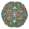
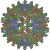
| Entry | Database: PDB / ID: 1qgt | ||||||
|---|---|---|---|---|---|---|---|
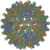
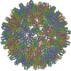
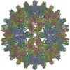
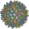
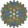
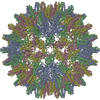
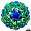
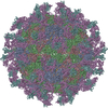
| Title | HUMAN HEPATITIS B VIRAL CAPSID (HBCAG) | ||||||
 Components Components | PROTEIN (HBV CAPSID PROTEIN) | ||||||
 Keywords Keywords | VIRUS / VIRAL CAPSID PROTEIN / Icosahedral virus | ||||||
| Function / homology |  Function and homology information Function and homology informationmicrotubule-dependent intracellular transport of viral material towards nucleus / T=4 icosahedral viral capsid / viral penetration into host nucleus / host cell / host cell cytoplasm / viral envelope / symbiont entry into host cell / structural molecule activity / DNA binding / RNA binding Similarity search - Function | ||||||
| Biological species |   Hepatitis B virus Hepatitis B virus | ||||||
| Method |  X-RAY DIFFRACTION / SYNCHROTRON / OTHER / Resolution: 3.3 Å X-RAY DIFFRACTION / SYNCHROTRON / OTHER / Resolution: 3.3 Å | ||||||
 Authors Authors | Leslie, A.G.W. / Wynne, S.A. / Crowther, R.A. | ||||||
 Citation Citation | Journal: Mol.Cell / Year: 1999 Title: The crystal structure of the human hepatitis B virus capsid. Authors: Wynne, S.A. / Crowther, R.A. / Leslie, A.G. #1: Journal: Acta Crystallogr.,Sect.D / Year: 1999Title: Crystallization of Hepatitis B Virus Core Protein Shells: Determination of Cryoprotectant Conditions and Preliminary X-Ray Characterization Authors: Wynne, S.A. / Leslie, A.G.W. / Butler, P.J.G. / Crowther, R.A. | ||||||
| History |
|
- Structure visualization
| Structure viewer | Molecule: MolmilJmol/JSmol |
|---|
- Downloads & links
-Download
| PDBx/mmCIF format | 1qgt.cif.gz | 113.5 KB | Display | PDBx/mmCIF format |
|---|---|---|---|---|
| PDB format | pdb1qgt.ent.gz | 90.7 KB | Display | PDB format |
| PDBx/mmJSON format | 1qgt.json.gz | Tree view | PDBx/mmJSON format | |
| Others |  Other downloads Other downloads |
-Validation report
| Arichive directory | https://data.pdbj.org/pub/pdb/validation_reports/qg/1qgtftp://data.pdbj.org/pub/pdb/validation_reports/qg/1qgt | HTTPS FTP |
|---|
-Related structure data
| Similar structure data |
|---|
-Links
 PDBj
PDBj

- Assembly
| Deposited unit | 
| |||||||||||||||||||||||||||||||||||||||||||||||||||||||||||||||||||||||||||||||||||||||||||||
|---|---|---|---|---|---|---|---|---|---|---|---|---|---|---|---|---|---|---|---|---|---|---|---|---|---|---|---|---|---|---|---|---|---|---|---|---|---|---|---|---|---|---|---|---|---|---|---|---|---|---|---|---|---|---|---|---|---|---|---|---|---|---|---|---|---|---|---|---|---|---|---|---|---|---|---|---|---|---|---|---|---|---|---|---|---|---|---|---|---|---|---|---|---|---|
| 1 | x 60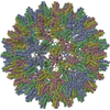
| |||||||||||||||||||||||||||||||||||||||||||||||||||||||||||||||||||||||||||||||||||||||||||||
| 2 |
| |||||||||||||||||||||||||||||||||||||||||||||||||||||||||||||||||||||||||||||||||||||||||||||
| 3 | x 5
| |||||||||||||||||||||||||||||||||||||||||||||||||||||||||||||||||||||||||||||||||||||||||||||
| 4 | x 6
| |||||||||||||||||||||||||||||||||||||||||||||||||||||||||||||||||||||||||||||||||||||||||||||
| 5 | 
| |||||||||||||||||||||||||||||||||||||||||||||||||||||||||||||||||||||||||||||||||||||||||||||
| 6 | x 30
| |||||||||||||||||||||||||||||||||||||||||||||||||||||||||||||||||||||||||||||||||||||||||||||
| Unit cell |
| |||||||||||||||||||||||||||||||||||||||||||||||||||||||||||||||||||||||||||||||||||||||||||||
| Symmetry | Point symmetry: (Hermann–Mauguin notation: 532 / Schoenflies symbol: I (icosahedral)) | |||||||||||||||||||||||||||||||||||||||||||||||||||||||||||||||||||||||||||||||||||||||||||||
| Noncrystallographic symmetry (NCS) | NCS oper:
|
-Components
| #1: Protein | Mass: 16866.283 Da / Num. of mol.: 4 / Fragment: ASSEMBLY DOMAIN Source method: isolated from a genetically manipulated source Source: (gene. exp.) Hepatitis B virus / Genus: Orthohepadnavirus / Strain: ISOLATED AT ST MARY'S HOSPITAL, LONDON / Description: THE CONSTRUCT WAS TRUNCATED AFTER RESIDUE 149 / Plasmid: PT7-SC / Species (production host): Escherichia coli / Production host:  Has protein modification | Y | |
|---|
-Experimental details
-Experiment
| Experiment | Method: X-RAY DIFFRACTION / Number of used crystals: 20 |
|---|
- Sample preparation
| Crystal | Density % sol: 82 % | ||||||||||||||||||||||||||||||||||||||||||||||||
|---|---|---|---|---|---|---|---|---|---|---|---|---|---|---|---|---|---|---|---|---|---|---|---|---|---|---|---|---|---|---|---|---|---|---|---|---|---|---|---|---|---|---|---|---|---|---|---|---|---|
| Crystal grow | pH: 6.5 Details: EQUAL VOLUMES OF PROTEIN (15MG/ML) IN 5MM TRIS-HCL, 150MM NACL PH7.5 AND 0.1M MES, PH 6.5, 0.1-0.4M (NH4)2SO4, 3.5-4% PEG20000 AND 20% BUTANEDIOL. HANGING DROP | ||||||||||||||||||||||||||||||||||||||||||||||||
| Crystal grow | *PLUS Temperature: 21 ℃ / Method: vapor diffusion, hanging dropDetails: drop consists of equal volume of protein and reservoir solutions | ||||||||||||||||||||||||||||||||||||||||||||||||
| Components of the solutions | *PLUS
|
-Data collection
| Diffraction | Mean temperature: 100 K |
|---|---|
| Diffraction source | Source: SYNCHROTRON / Site: ESRF  / Beamline: ID2 / Wavelength: 0.99188 / Beamline: ID2 / Wavelength: 0.99188 |
| Detector | Type: MAR scanner 345 mm plate / Detector: IMAGE PLATE / Date: Jan 1, 1998 |
| Radiation | Protocol: SINGLE WAVELENGTH / Monochromatic (M) / Laue (L): M / Scattering type: x-ray |
| Radiation wavelength | Wavelength: 0.99188 Å / Relative weight: 1 |
| Reflection | Resolution: 3.3→38.8 Å / Num. obs: 727106 / % possible obs: 94.9 % / Redundancy: 2.9 % / Biso Wilson estimate: 59 Å2 / Rmerge(I) obs: 0.153 / Net I/σ(I): 6.4 |
| Reflection shell | Resolution: 3.3→3.48 Å / Redundancy: 2 % / Rmerge(I) obs: 0.782 / Mean I/σ(I) obs: 0.9 / % possible all: 78.2 |
| Reflection shell | *PLUS % possible obs: 78.2 % |
- Processing
| Software |
| ||||||||||||||||||||||||||||||||||||||||||||||||||||||||||||
|---|---|---|---|---|---|---|---|---|---|---|---|---|---|---|---|---|---|---|---|---|---|---|---|---|---|---|---|---|---|---|---|---|---|---|---|---|---|---|---|---|---|---|---|---|---|---|---|---|---|---|---|---|---|---|---|---|---|---|---|---|---|
| Refinement | Method to determine structure: OTHER / Resolution: 3.3→8 Å / Data cutoff high absF: 0 / Data cutoff low absF: 0 / Isotropic thermal model: GROUP B'S / σ(F): 0 / Details: STRICT ICOSAHEDRAL SYMMETRY IMPOSED
| ||||||||||||||||||||||||||||||||||||||||||||||||||||||||||||
| Displacement parameters | Biso mean: 54 Å2 | ||||||||||||||||||||||||||||||||||||||||||||||||||||||||||||
| Refinement step | Cycle: LAST / Resolution: 3.3→8 Å
| ||||||||||||||||||||||||||||||||||||||||||||||||||||||||||||
| Refine LS restraints |
| ||||||||||||||||||||||||||||||||||||||||||||||||||||||||||||
| Refine LS restraints NCS | NCS model details: CONSTRAINTS | ||||||||||||||||||||||||||||||||||||||||||||||||||||||||||||
| LS refinement shell | Resolution: 3.3→3.44 Å / Total num. of bins used: 8 /
| ||||||||||||||||||||||||||||||||||||||||||||||||||||||||||||
| Xplor file | Serial no: 1 / Param file: PARHCSDX.PRO / Topol file: TOPHCSDX.PRO | ||||||||||||||||||||||||||||||||||||||||||||||||||||||||||||
| Software | *PLUS Name: X-PLOR / Version: 3.851 / Classification: refinement | ||||||||||||||||||||||||||||||||||||||||||||||||||||||||||||
| Refinement | *PLUS Rfactor obs: 0.271 | ||||||||||||||||||||||||||||||||||||||||||||||||||||||||||||
| Solvent computation | *PLUS | ||||||||||||||||||||||||||||||||||||||||||||||||||||||||||||
| Displacement parameters | *PLUS | ||||||||||||||||||||||||||||||||||||||||||||||||||||||||||||
| Refine LS restraints | *PLUS
|
