 Movie
Movie Controller


+ Open data

- Basic information
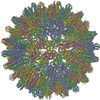
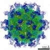
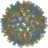
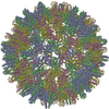
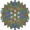
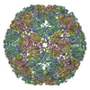
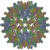
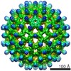
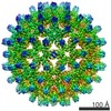
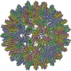
| Entry | Database: PDB / ID: 6hu4 | ||||||
|---|---|---|---|---|---|---|---|
| Title | F97L Hepatitis B core protein capsid | ||||||
 Components Components | Capsid protein | ||||||
 Keywords Keywords | VIRUS LIKE PARTICLE / Hapatitis B core protein Hbc Hbcag / VIRUS LIKE PARTICLE premature envelopment mutant F97L | ||||||
| Function / homology |  Function and homology information Function and homology informationmicrotubule-dependent intracellular transport of viral material towards nucleus / T=4 icosahedral viral capsid / viral penetration into host nucleus / host cell / host cell cytoplasm / symbiont entry into host cell / structural molecule activity / DNA binding / RNA binding Similarity search - Function | ||||||
| Biological species |   Hepatitis B virus Hepatitis B virus | ||||||
| Method | ELECTRON MICROSCOPY / single particle reconstruction / cryo EM / Resolution: 2.64 Å | ||||||
 Authors Authors | Bottcher, B. / Nassal, M. | ||||||
| Funding support |  Germany, 1items Germany, 1items
| ||||||
 Citation Citation | Journal: J Mol Biol / Year: 2018 Title: Structure of Mutant Hepatitis B Core Protein Capsids with Premature Secretion Phenotype. Authors: Bettina Böttcher / Michael Nassal / Abstract: Hepatitis B virus is a major human pathogen that consists of a viral genome surrounded by an icosahedrally ordered core protein and a polymorphic, lipidic envelope that is densely packed with surface ...Hepatitis B virus is a major human pathogen that consists of a viral genome surrounded by an icosahedrally ordered core protein and a polymorphic, lipidic envelope that is densely packed with surface proteins. A point mutation in the core protein in which a phenylalanine at position 97 is exchanged for a smaller leucine leads to premature envelopment of the capsid before the genome maturation is fully completed. We have used electron cryo-microscopy and image processing to investigate how the point mutation affects the structure of the capsid at 2.6- to 2.8 Å-resolution. We found that in the mutant the smaller side chain at position 97 is displaced, increasing the size of an adjacent pocket in the center of the spikes of the capsid. In the mutant, this pocket is filled with an unknown density. Phosphorylation of serine residues in the unresolved C-terminal domain of the mutant leaves the structure of the ordered capsid largely unchanged. However, we were able to resolve several previously unresolved residues downstream of proline 144 that precede the phosphorylation-sites. These residues pack against the neighboring subunits and increase the inter-dimer contact suggesting that the C-termini play an important role in capsid stabilization and provide a much larger interaction interface than previously observed. | ||||||
| History |
|
- Structure visualization
| Movie |
Movie viewer |
|---|---|
| Structure viewer | Molecule: MolmilJmol/JSmol |
- Downloads & links
-Download
| PDBx/mmCIF format | 6hu4.cif.gz | 142.4 KB | Display | PDBx/mmCIF format |
|---|---|---|---|---|
| PDB format | pdb6hu4.ent.gz | 107.2 KB | Display | PDB format |
| PDBx/mmJSON format | 6hu4.json.gz | Tree view | PDBx/mmJSON format | |
| Others |  Other downloads Other downloads |
-Validation report
| Arichive directory | https://data.pdbj.org/pub/pdb/validation_reports/hu/6hu4ftp://data.pdbj.org/pub/pdb/validation_reports/hu/6hu4 | HTTPS FTP |
|---|
-Related structure data
| Related structure data |  0272MC 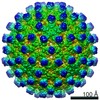 0271C 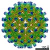 0273C 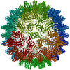 6htxC 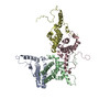 6hu7C C: citing same article ( M: map data used to model this data |
|---|---|
| Similar structure data |
-Links
 PDBj
PDBj

- Assembly
| Deposited unit | 
|
|---|---|
| 1 | x 60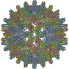
|
| 2 |
|
| 3 | x 5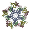
|
| 4 | x 6
|
| 5 | 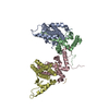
|
| Symmetry | Point symmetry: (Schoenflies symbol: I (icosahedral)) |
-Components
| #1: Protein | Mass: 21112.199 Da / Num. of mol.: 8 Source method: isolated from a genetically manipulated source Details: Hepatitis B core protein premature envelopment mutant F97L Source: (gene. exp.) Hepatitis B virus / Production host:  Has protein modification | Y | |
|---|
-Experimental details
-Experiment
| Experiment | Method: ELECTRON MICROSCOPY |
|---|---|
| EM experiment | Aggregation state: PARTICLE / 3D reconstruction method: single particle reconstruction |
- Sample preparation
| Component | Name: Hepatitis B virus / Type: VIRUS / Entity ID: all / Source: RECOMBINANT | |||||||||||||||
|---|---|---|---|---|---|---|---|---|---|---|---|---|---|---|---|---|
| Molecular weight | Value: 4.8 MDa / Experimental value: NO | |||||||||||||||
| Source (natural) | Organism: Hepatitis B virus | |||||||||||||||
| Source (recombinant) | Organism: | |||||||||||||||
| Details of virus | Empty: NO / Enveloped: NO / Isolate: STRAIN / Type: VIRUS-LIKE PARTICLE | |||||||||||||||
| Natural host | Organism: Hepatitis B virus / Strain: ayw | |||||||||||||||
| Virus shell | Name: WT Hbc / Diameter: 340 nm / Triangulation number (T number): 4 | |||||||||||||||
| Buffer solution | pH: 7.7 / Details: 25 mM Tris pH 7.7, 150 mM NaCl | |||||||||||||||
| Buffer component |
| |||||||||||||||
| Specimen | Conc.: 5 mg/ml / Embedding applied: NO / Shadowing applied: NO / Staining applied: NO / Vitrification applied: YES Details: sample is purified from e.coli via sucrose density gradient. sucrose is removed via buffer exchange on a concentrator | |||||||||||||||
| Specimen support | Grid material: GOLD / Grid mesh size: 300 divisions/in. / Grid type: Quantifoil, UltrAuFoil, R1.2/1.3 | |||||||||||||||
| Vitrification | Instrument: FEI VITROBOT MARK IV / Cryogen name: ETHANE / Humidity: 100 % / Chamber temperature: 4 K Details: Samples were vitrified with an FEI Vitrobot IV, which was operated at 100% humidity at 4C. 2.5-3.5 ul of sample were applied to grids (Quantifoil UltrAuFoil R1.3/1.2 300 mesh gold grids ) ...Details: Samples were vitrified with an FEI Vitrobot IV, which was operated at 100% humidity at 4C. 2.5-3.5 ul of sample were applied to grids (Quantifoil UltrAuFoil R1.3/1.2 300 mesh gold grids ) that were glow discharged in air for 1-2 min. The sample was incubated for 30 s before blotting for 3 s with a blot force between 0 and 5. |
- Electron microscopy imaging
| Experimental equipment |  Model: Titan Krios / Image courtesy: FEI Company |
|---|---|
| Microscopy | Model: FEI TITAN KRIOS |
| Electron gun | Electron source:  FIELD EMISSION GUN / Accelerating voltage: 300 kV / Illumination mode: FLOOD BEAM FIELD EMISSION GUN / Accelerating voltage: 300 kV / Illumination mode: FLOOD BEAM |
| Electron lens | Mode: BRIGHT FIELD / Nominal magnification: 75000 X / Calibrated defocus min: 370 nm / Calibrated defocus max: 2560 nm / Cs: 2.7 mm / C2 aperture diameter: 70 µm / Alignment procedure: COMA FREE |
| Specimen holder | Cryogen: NITROGEN / Specimen holder model: FEI TITAN KRIOS AUTOGRID HOLDER |
| Image recording | Electron dose: 28 e/Å2 / Detector mode: COUNTING / Film or detector model: GATAN K2 SUMMIT (4k x 4k) / Num. of grids imaged: 1 / Num. of real images: 1948 |
| EM imaging optics | Energyfilter name: GIF Quantum LS / Energyfilter slit width: 20 eV |
| Image scans | Movie frames/image: 28 / Used frames/image: 1-28 |
- Processing
| Software | Name: PHENIX / Version: 1.10.1_2155: / Classification: refinement | |||||||||||||||||||||||||||||||||||||||||||||||||||||||||||||||||
|---|---|---|---|---|---|---|---|---|---|---|---|---|---|---|---|---|---|---|---|---|---|---|---|---|---|---|---|---|---|---|---|---|---|---|---|---|---|---|---|---|---|---|---|---|---|---|---|---|---|---|---|---|---|---|---|---|---|---|---|---|---|---|---|---|---|---|
| EM software |
| |||||||||||||||||||||||||||||||||||||||||||||||||||||||||||||||||
| CTF correction | Type: PHASE FLIPPING AND AMPLITUDE CORRECTION | |||||||||||||||||||||||||||||||||||||||||||||||||||||||||||||||||
| Particle selection | Num. of particles selected: 82798 / Details: template based auto picking | |||||||||||||||||||||||||||||||||||||||||||||||||||||||||||||||||
| Symmetry | Point symmetry: I (icosahedral) | |||||||||||||||||||||||||||||||||||||||||||||||||||||||||||||||||
| 3D reconstruction | Resolution: 2.64 Å / Resolution method: FSC 0.143 CUT-OFF / Num. of particles: 38292 / Algorithm: FOURIER SPACE / Num. of class averages: 1 / Symmetry type: POINT | |||||||||||||||||||||||||||||||||||||||||||||||||||||||||||||||||
| Atomic model building | B value: 85 / Protocol: FLEXIBLE FIT / Space: REAL | |||||||||||||||||||||||||||||||||||||||||||||||||||||||||||||||||
| Atomic model building | PDB-ID: 6HTX Accession code: 6HTX / Source name: PDB / Type: experimental model | |||||||||||||||||||||||||||||||||||||||||||||||||||||||||||||||||
| Refine LS restraints |
|
