 Movie
Movie Controller


+ Open data

- Basic information
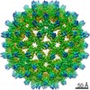
| Entry | Database: EMDB / ID: EMD-21495 | |||||||||
|---|---|---|---|---|---|---|---|---|---|---|
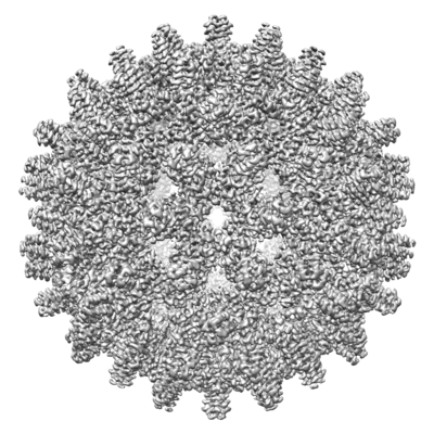
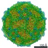
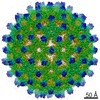
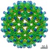
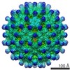
| Title | CryoEM density map of wild type HBV capsid | |||||||||
 Map data Map data | CryoEm density map of wild type HBV capsid | |||||||||
 Sample Sample |
| |||||||||
 Keywords Keywords | HBV / Core protein / VIRUS | |||||||||
| Function / homology |  Function and homology information Function and homology informationmicrotubule-dependent intracellular transport of viral material towards nucleus / T=4 icosahedral viral capsid / viral penetration into host nucleus / host cell / host cell cytoplasm / symbiont entry into host cell / structural molecule activity / DNA binding / RNA binding / identical protein binding Similarity search - Function | |||||||||
| Biological species |  Hepatitis B virus genotype D subtype adw (isolate United Kingdom/adyw/1979) Hepatitis B virus genotype D subtype adw (isolate United Kingdom/adyw/1979) | |||||||||
| Method | single particle reconstruction / cryo EM / Resolution: 3.6 Å | |||||||||
 Authors Authors | Zhao Z / Wang J | |||||||||
| Funding support |  United States, 1 items United States, 1 items
| |||||||||
 Citation Citation | Journal: ACS Chem Biol / Year: 2020 Title: The Integrity of the Intradimer Interface of the Hepatitis B Virus Capsid Protein Dimer Regulates Capsid Self-Assembly. Authors: Zhongchao Zhao / Joseph Che-Yen Wang / Carolina Pérez Segura / Jodi A Hadden-Perilla / Adam Zlotnick / Abstract: During the hepatitis B virus lifecycle, 120 copies of homodimeric capsid protein assemble around a copy of reverse transcriptase and viral RNA and go on to produce an infectious virion. Assembly ...During the hepatitis B virus lifecycle, 120 copies of homodimeric capsid protein assemble around a copy of reverse transcriptase and viral RNA and go on to produce an infectious virion. Assembly needs to be tightly regulated by protein conformational change to ensure symmetry, fidelity, and reproducibility. Here, we show that structures at the intradimer interface regulate conformational changes at the distal interdimer interface and so regulate assembly. A pair of interacting charged residues, D78 from each monomer, conspicuously located at the top of a four-helix bundle that forms the intradimer interface, were mutated to serine to disrupt communication between the two monomers. The mutation slowed assembly and destabilized the dimer to thermal and chemical denaturation. Mutant dimers showed evidence of transient partial unfolding based on the appearance of new proteolytically sensitive sites. Though the mutant dimer was less stable, the resulting capsids were as stable as the wildtype, based on assembly and thermal denaturation studies. Cryo-EM image reconstructions of capsid indicated that the subunits adopted an "open" state more usually associated with a free dimer and that the spike tips were either disordered or highly flexible. Molecular dynamics simulations provide mechanistic explanations for these results, suggesting that D78 stabilizes helix 4a, which forms part of the intradimer interface, by capping its N-terminus and hydrogen-bonding to nearby residues, whereas the D78S mutation disrupts these interactions, leading to partial unwinding of helix 4a. This in turn weakens the connection from helix 4 and the intradimer interface to helix 5, which forms the interdimer interface. | |||||||||
| History |
|
- Structure visualization
| Movie |
Movie viewer |
|---|---|
| Structure viewer | EM map: SurfViewMolmilJmol/JSmol |
| Supplemental images |
- Downloads & links
-EMDB archive
| Map data | emd_21495.map.gz | 62.6 MB | EMDB map data format | |
|---|---|---|---|---|
| Header (meta data) | emd-21495-v30.xmlemd-21495.xml | 9.8 KB 9.8 KB | Display Display | EMDB header |
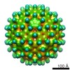
| Images |  emd_21495.png emd_21495.png | 186.5 KB | ||
| Filedesc metadata | emd-21495.cif.gz | 4.9 KB | ||
| Archive directory |  http://ftp.pdbj.org/pub/emdb/structures/EMD-21495ftp://ftp.pdbj.org/pub/emdb/structures/EMD-21495 http://ftp.pdbj.org/pub/emdb/structures/EMD-21495ftp://ftp.pdbj.org/pub/emdb/structures/EMD-21495 | HTTPS FTP |
-Related structure data
| Related structure data |  6vzpMC  6w0kC C: citing same article ( M: atomic model generated by this map |
|---|---|
| Similar structure data |


-Links
| EMDB pages | EMDB (EBI/PDBe) / EMDataResource |
|---|---|
| Related items in Molecule of the Month |


-Map
| File | Download / File: emd_21495.map.gz / Format: CCP4 / Size: 476.8 MB / Type: IMAGE STORED AS FLOATING POINT NUMBER (4 BYTES) | ||||||||||||||||||||||||||||||||||||||||||||||||||||||||||||||||||||
|---|---|---|---|---|---|---|---|---|---|---|---|---|---|---|---|---|---|---|---|---|---|---|---|---|---|---|---|---|---|---|---|---|---|---|---|---|---|---|---|---|---|---|---|---|---|---|---|---|---|---|---|---|---|---|---|---|---|---|---|---|---|---|---|---|---|---|---|---|---|
| Annotation | CryoEm density map of wild type HBV capsid | ||||||||||||||||||||||||||||||||||||||||||||||||||||||||||||||||||||
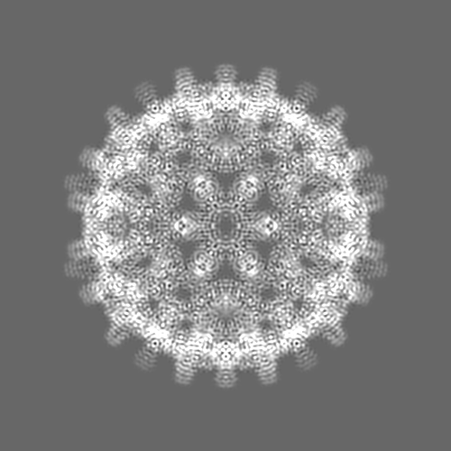
| Projections & slices | Image control
Images are generated by Spider. | ||||||||||||||||||||||||||||||||||||||||||||||||||||||||||||||||||||
| Voxel size | X=Y=Z: 0.96 Å | ||||||||||||||||||||||||||||||||||||||||||||||||||||||||||||||||||||
| Density |
| ||||||||||||||||||||||||||||||||||||||||||||||||||||||||||||||||||||
| Symmetry | Space group: 1 | ||||||||||||||||||||||||||||||||||||||||||||||||||||||||||||||||||||
| Details | EMDB XML:
CCP4 map header:
| ||||||||||||||||||||||||||||||||||||||||||||||||||||||||||||||||||||
 Z (Sec.)
 Y (Row.)
 X (Col.)


-Supplemental data
- Sample components
-Entire : HBV wild type capsid
| Entire | Name: HBV wild type capsid |
|---|---|
| Components |
|
-Supramolecule #1: HBV wild type capsid
| Supramolecule | Name: HBV wild type capsid / type: complex / ID: 1 / Parent: 0 / Macromolecule list: all |
|---|---|
| Source (natural) | Organism: Hepatitis B virus genotype D subtype adw (isolate United Kingdom/adyw/1979) Strain: isolate United Kingdom/adyw/1979 |
-Macromolecule #1: Capsid protein
| Macromolecule | Name: Capsid protein / type: protein_or_peptide / ID: 1 / Number of copies: 4 / Enantiomer: LEVO |
|---|---|
| Source (natural) | Organism: Hepatitis B virus genotype D subtype adw (isolate United Kingdom/adyw/1979) Strain: isolate United Kingdom/adyw/1979 |
| Molecular weight | Theoretical: 16.784158 KDa |
| Recombinant expression | Organism:  |
| Sequence | String: MDIDPYKEFG ATVELLSFLP SDFFPSVRDL LDTAAALYRD ALESPEHCSP HHTALRQAIL CWGDLMTLAT WVGTNLEDPA SRDLVVSYV NTNVGLKFRQ LLWFHISCLT FGRETVLEYL VSFGVWIRTP PAYRPPNAPI LSTLPETTVV UniProtKB: Capsid protein |
-Experimental details
-Structure determination
| Method | cryo EM |
|---|---|
 Processing Processing | single particle reconstruction |
| Aggregation state | particle |
-Sample preparation
| Concentration | 15 mg/mL |
|---|---|
| Buffer | pH: 7.5 |
| Grid | Details: unspecified |
| Vitrification | Cryogen name: ETHANE |
- Electron microscopy
| Microscope | FEI TALOS ARCTICA |
|---|---|
| Image recording | Film or detector model: FEI FALCON III (4k x 4k) / Average electron dose: 30.0 e/Å2 |
| Electron beam | Acceleration voltage: 200 kV / Electron source:  FIELD EMISSION GUN FIELD EMISSION GUN |
| Electron optics | Illumination mode: FLOOD BEAM / Imaging mode: BRIGHT FIELD |
| Experimental equipment |  Model: Talos Arctica / Image courtesy: FEI Company |
-Image processing
| Startup model | Type of model: PDB ENTRY PDB model - PDB ID: |
|---|---|
| Final reconstruction | Resolution.type: BY AUTHOR / Resolution: 3.6 Å / Resolution method: FSC 0.143 CUT-OFF / Number images used: 139740 |
| Initial angle assignment | Type: RANDOM ASSIGNMENT |
| Final angle assignment | Type: PROJECTION MATCHING |

-Atomic model buiding 1
| Refinement | Protocol: RIGID BODY FIT |
|---|---|
| Output model | PDB-6vzp: |
