 Movie
Movie Controller


+ Open data

- Basic information
| Entry | Database: PDB / ID: 6q81 | ||||||
|---|---|---|---|---|---|---|---|
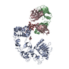
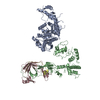
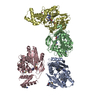
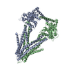
| Title | Structure of P-glycoprotein(ABCB1) in the post-hydrolytic state | ||||||
 Components Components | P-glycoprotein (ABCB1) | ||||||
 Keywords Keywords | MEMBRANE PROTEIN / P-glycoprotein / ABCB1 / ATP-binding cassette / transporter / protein structure | ||||||
| Function / homology |  Function and homology information Function and homology informationAtorvastatin ADME / aldosterone secretion / neural tissue regeneration / Prednisone ADME / cellular response to nonylphenol / cellular response to borneol / response to codeine / cellular response to mycotoxin / daunorubicin transport / positive regulation of response to drug ...Atorvastatin ADME / aldosterone secretion / neural tissue regeneration / Prednisone ADME / cellular response to nonylphenol / cellular response to borneol / response to codeine / cellular response to mycotoxin / daunorubicin transport / positive regulation of response to drug / terpenoid transport / ceramide floppase activity / regulation of intestinal absorption / response to cyclosporin A / cellular response to external biotic stimulus / response to antineoplastic agent / positive regulation of establishment of Sertoli cell barrier / negative regulation of sensory perception of pain / carboxylic acid transmembrane transport / floppase activity / ceramide translocation / response to quercetin / carboxylic acid transmembrane transporter activity / ABC-family protein mediated transport / establishment of blood-retinal barrier / phosphatidylethanolamine flippase activity / protein localization to bicellular tight junction / phosphatidylcholine floppase activity / cardiac muscle cell differentiation / response to thyroxine / xenobiotic transport across blood-brain barrier / establishment of blood-brain barrier / intercellular canaliculus / xenobiotic detoxification by transmembrane export across the plasma membrane / export across plasma membrane / P-type phospholipid transporter / transepithelial transport / cellular response to L-glutamate / response to vitamin A / adult heart development / ABC-type xenobiotic transporter / response to glucagon / response to vitamin D / intestinal absorption / response to alcohol / response to glycoside / phospholipid translocation / ABC-type xenobiotic transporter activity / cellular hyperosmotic salinity response / cellular response to alkaloid / female gonad development / exploration behavior / cellular response to antibiotic / maintenance of blood-brain barrier / efflux transmembrane transporter activity / ATPase-coupled transmembrane transporter activity / xenobiotic transmembrane transporter activity / cellular response to dexamethasone stimulus / response to cadmium ion / xenobiotic transport / transmembrane transporter activity / response to progesterone / lactation / stem cell proliferation / neurogenesis / placenta development / regulation of chloride transport / proteasomal protein catabolic process / cellular response to estradiol stimulus / brush border membrane / female pregnancy / circadian rhythm / cellular response to tumor necrosis factor / G2/M transition of mitotic cell cycle / response to toxic substance / epidermal growth factor receptor signaling pathway / heart development / cellular response to lipopolysaccharide / gene expression / response to hypoxia / apical plasma membrane / response to xenobiotic stimulus / ubiquitin protein ligase binding / cell surface / ATP hydrolysis activity / ATP binding / membrane / plasma membrane / cytoplasm Similarity search - Function | ||||||
| Biological species |  | ||||||
| Method | ELECTRON MICROSCOPY / single particle reconstruction / cryo EM / Resolution: 7.9 Å | ||||||
 Authors Authors | Ford, R.C. / Thonghin, N. / Collins, R.F. / Barbieri, A. / Shafi, T. / Siebert, A. | ||||||
 Citation Citation | Journal: BMC Struct Biol / Year: 2018 Title: Novel features in the structure of P-glycoprotein (ABCB1) in the post-hydrolytic state as determined at 7.9 Å resolution. Authors: Nopnithi Thonghin / Richard F Collins / Alessandro Barbieri / Talha Shafi / Alistair Siebert / Robert C Ford /  Abstract: BACKGROUND: P-glycoprotein (ABCB1) is an ATP-binding cassette transporter that plays an important role in the clearance of drugs and xenobiotics and is associated with multi-drug resistance in cancer. ...BACKGROUND: P-glycoprotein (ABCB1) is an ATP-binding cassette transporter that plays an important role in the clearance of drugs and xenobiotics and is associated with multi-drug resistance in cancer. Although several P-glycoprotein structures are available, these are either at low resolution, or represent mutated and/or quiescent states of the protein. RESULTS: In the post-hydrolytic state the structure of the wild-type protein has been resolved at about 8 Å resolution. The cytosolic nucleotide-binding domains (NBDs) are separated but ADP ...RESULTS: In the post-hydrolytic state the structure of the wild-type protein has been resolved at about 8 Å resolution. The cytosolic nucleotide-binding domains (NBDs) are separated but ADP remains bound, especially at the first NBD. Gaps in the transmembrane domains (TMDs) that connect to an inner hydrophilic cavity are filled by density emerging from the annular detergent micelle. The NBD-TMD linker is partly resolved, being located between the NBDs and close to the Signature regions involved in cooperative NBD dimerization. This, and the gap-filling detergent suggest steric impediment to NBD dimerization in the post-hydrolytic state. Two central regions of density lie in two predicted drug-binding sites, implying that the protein may adventitiously bind hydrophobic substances even in the post-hydrolytic state. The previously unresolved N-terminal extension was observed, and the data suggests these 30 residues interact with the headgroup region of the lipid bilayer. CONCLUSION: The structural data imply that (i) a low basal ATPase activity is ensured by steric blockers of NBD dimerization and (ii) allocrite access to the central cavity may be structurally linked ...CONCLUSION: The structural data imply that (i) a low basal ATPase activity is ensured by steric blockers of NBD dimerization and (ii) allocrite access to the central cavity may be structurally linked to NBD dimerization, giving insights into the mechanism of drug-stimulation of P-glycoprotein activity. | ||||||
| History |
|
- Structure visualization
| Movie |
Movie viewer |
|---|---|
| Structure viewer | Molecule: MolmilJmol/JSmol |
- Downloads & links
-Download
| PDBx/mmCIF format | 6q81.cif.gz | 214.1 KB | Display | PDBx/mmCIF format |
|---|---|---|---|---|
| PDB format | pdb6q81.ent.gz | 161 KB | Display | PDB format |
| PDBx/mmJSON format | 6q81.json.gz | Tree view | PDBx/mmJSON format | |
| Others |  Other downloads Other downloads |
-Validation report
| Arichive directory | https://data.pdbj.org/pub/pdb/validation_reports/q8/6q81ftp://data.pdbj.org/pub/pdb/validation_reports/q8/6q81 | HTTPS FTP |
|---|
-Related structure data
| Related structure data | 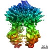 4391MC  6gdiC M: map data used to model this data C: citing same article ( |
|---|---|
| Similar structure data |


-Links
 PDBj
PDBj


- Assembly
| Deposited unit | 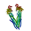
|
|---|---|
| 1 |
|
-Components
| #1: Protein | Mass: 140806.766 Da / Num. of mol.: 1 Source method: isolated from a genetically manipulated source Source: (gene. exp.)  |
|---|---|
| #2: Chemical |   Mass: 427.201 Da / Num. of mol.: 2 / Source method: obtained synthetically / Formula: C10H15N5O10P2 / Comment: ADP, energy-carrying molecule*YM Mass: 427.201 Da / Num. of mol.: 2 / Source method: obtained synthetically / Formula: C10H15N5O10P2 / Comment: ADP, energy-carrying molecule*YM |
-Experimental details
-Experiment
| Experiment | Method: ELECTRON MICROSCOPY |
|---|---|
| EM experiment | Aggregation state: PARTICLE / 3D reconstruction method: single particle reconstruction |
- Sample preparation
| Component | Name: P-glycoprotein trapped in the post-hydrolytic state. / Type: ORGANELLE OR CELLULAR COMPONENT / Details: ATP and Vanadate added / Entity ID: #1 / Source: RECOMBINANT |
|---|---|
| Molecular weight | Value: 142 kDa/nm / Experimental value: NO |
| Source (natural) | Organism: |
| Source (recombinant) | Organism: |
| Buffer solution | pH: 8 |
| Specimen | Embedding applied: NO / Shadowing applied: NO / Staining applied: NO / Vitrification applied: YES |
| Vitrification | Cryogen name: ETHANE |
- Electron microscopy imaging
| Experimental equipment |  Model: Titan Krios / Image courtesy: FEI Company |
|---|---|
| Microscopy | Model: FEI TITAN KRIOS |
| Electron gun | Electron source:  FIELD EMISSION GUN / Accelerating voltage: 300 kV / Illumination mode: FLOOD BEAM FIELD EMISSION GUN / Accelerating voltage: 300 kV / Illumination mode: FLOOD BEAM |
| Electron lens | Mode: BRIGHT FIELD |
| Image recording | Electron dose: 70 e/Å2 / Film or detector model: GATAN K2 SUMMIT (4k x 4k) |
- Processing
| EM software |
| ||||||||||||
|---|---|---|---|---|---|---|---|---|---|---|---|---|---|
| CTF correction | Type: PHASE FLIPPING ONLY | ||||||||||||
| Symmetry | Point symmetry: C1 (asymmetric) | ||||||||||||
| 3D reconstruction | Method: SINGLE PARTICLE / Resolution: 7.9 Å / Resolution method: FSC 0.143 CUT-OFF / Num. of particles: 135357 / Symmetry type: POINT |
